Explore
 Validate
 Learn
Learn Western blot
Antibody data
- Antibody Data
- Antigen structure
- References [1]
- Comments [0]
- Validations
- Western blot [2]
- Immunocytochemistry [1]
- Immunohistochemistry [1]
Submit
Validation data
Reference
Comment
Report error
- Product number
- MAB6405 - Provider product page

- Provider
- R&D Systems
- Product name
- Human Bad Antibody
- Antibody type
- Monoclonal
- Description
- Protein A or G purified from hybridoma culture supernatant. Detects human Bad in direct ELISAs. In direct ELISAs, no cross-reactivity with recombinant mouse (rm) Bad (aa 1-130), rmBcl-2 alpha , recombinant human (rh) Bcl-2, or rhBCL2L12 is observed.
- Reactivity
- Human
- Host
- Mouse
- Conjugate
- Unconjugated
- Antigen sequence
Q92934- Isotype
- IgG
- Antibody clone number
- 612312
- Vial size
- 100 ug
- Concentration
- LYOPH
- Storage
- Use a manual defrost freezer and avoid repeated freeze-thaw cycles. 12 months from date of receipt, -20 to -70 °C as supplied. 1 month, 2 to 8 °C under sterile conditions after reconstitution. 6 months, -20 to -70 °C under sterile conditions after reconstitution.
Submitted references FK506 Attenuates the MRP1-Mediated Chemoresistant Phenotype in Glioblastoma Stem-Like Cells.
Torres Á, Arriagada V, Erices JI, Toro MLÁ, Rocha JD, Niechi I, Carrasco C, Oyarzún C, Quezada C
International journal of molecular sciences 2018 Sep 11;19(9)
International journal of molecular sciences 2018 Sep 11;19(9)
No comments: Submit comment
Supportive validation
- Submitted by
- R&D Systems (provider)
- Main image
- Experimental details
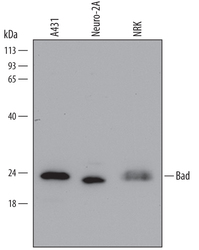
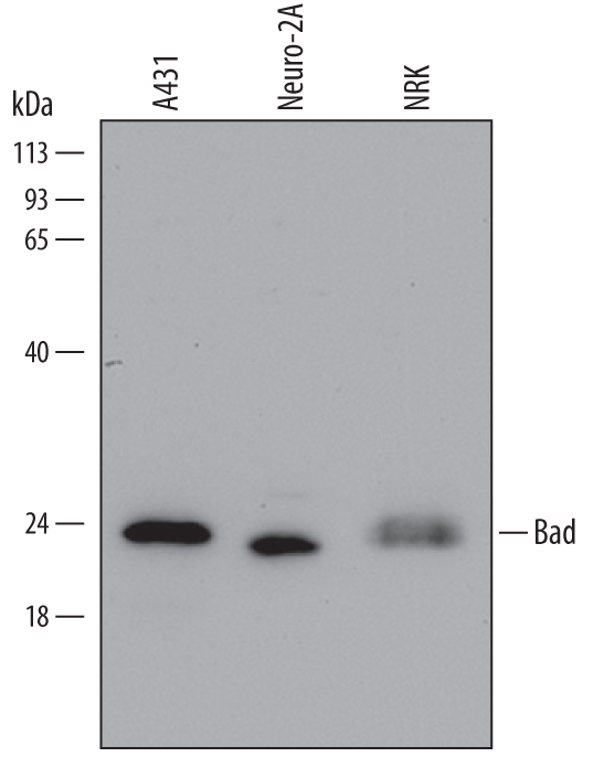
- Detection of Human, Mouse, and Rat Bad by Western Blot. Western blot shows lysates of A431 human epithelial carcinoma cell line, Neuro-2A mouse neuroblastoma cell line, and NRK rat normal kidney cell line. PVDF Membrane was probed with 0.5 µg/mL of Mouse Anti-Human/Mouse/Rat Bad Monoclonal Antibody (Catalog # MAB6405) followed by HRP-conjugated Anti-Mouse IgG Secondary Antibody (Catalog # HAF007). A specific band was detected for Bad at approximately 23 kDa (as indicated). This experiment was conducted under reducing conditions and using Immunoblot Buffer Group 2.
- Submitted by
- R&D Systems (provider)
- Main image
- Experimental details
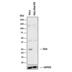
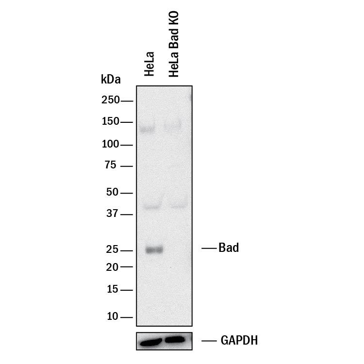
- Western Blot Shows Human Bad Specificity by Using Knockout Cell Line. Western blot shows lysates of HeLa human cervical epithelial carcinoma parental cell line and Bad knockout HeLa cell line (KO). PVDF membrane was probed with 0.5 µg/mL of Mouse Anti-Human Bad Monoclonal Antibody (Catalog # MAB6405) followed by HRP-conjugated Anti-Mouse IgG Secondary Antibody (Catalog # HAF018). A specific band was detected for Bad at approximately 25 kDa (as indicated) in the parental HeLa cell line, but is not detectable in knockout HeLa cell line. GAPDH (Catalog # MAB5718) is shown as a loading control. This experiment was conducted under reducing conditions and using Immunoblot Buffer Group 1.
Supportive validation
- Submitted by
- R&D Systems (provider)
- Main image
- Experimental details
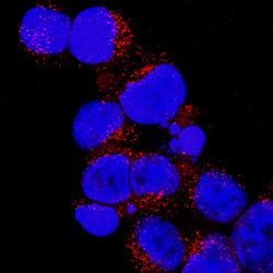
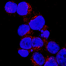
- Bad in Jurkat Human Cell Line. Bad was detected in immersion fixed Jurkat human acute T cell leukemia cell line using Mouse Anti-Human Bad Monoclonal Antibody (Catalog # MAB6405) at 25 µg/mL for 3 hours at room temperature. Cells were stained using the NorthernLights™ 557-conjugated Anti-Mouse IgG Secondary Antibody (red; Catalog # NL007) and counterstained with DAPI (blue). Specific staining was localized to cytoplasm. View our protocol for Fluorescent ICC Staining of Non-adherent Cells.
Supportive validation
- Submitted by
- R&D Systems (provider)
- Main image
- Experimental details
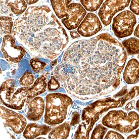
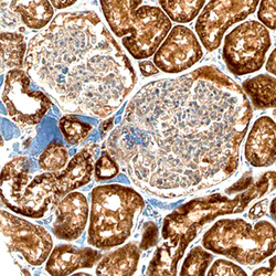
- Bad in Human Kidney. Bad was detected in immersion fixed paraffin-embedded sections of human kidney using Mouse Anti-Human/Mouse/Rat Bad Monoclonal Antibody (Catalog # MAB6405) at 15 µg/mL overnight at 4 °C. Before incubation with the primary antibody, tissue was subjected to heat-induced epitope retrieval using Antigen Retrieval Reagent-Basic (Catalog # CTS013). Tissue was stained using the Anti-Mouse HRP-DAB Cell & Tissue Staining Kit (brown; Catalog # CTS002) and counterstained with hematoxylin (blue). Specific staining was localized to cytoplasm of epithelial cells in convoluted tubules. View our protocol for Chromogenic IHC Staining of Paraffin-embedded Tissue Sections.