 Explore
 Validate
 Learn
Learn Western blot
Antibody data
- Antibody Data
- Antigen structure
- References [6]
- Comments [0]
- Validations
- Western blot [2]
- Immunocytochemistry [1]
- Immunohistochemistry [2]
Submit
Validation data
Reference
Comment
Report error
- Product number
- AF820 - Provider product page

- Provider
- R&D Systems
- Product name
- Human/Mouse Bax Antibody
- Antibody type
- Polyclonal
- Description
- Antigen Affinity-purified. Detects human and mouse Bax.
- Reactivity
- Human, Mouse
- Host
- Rabbit
- Conjugate
- Unconjugated
- Isotype
- IgG
- Vial size
- 30 ug
- Concentration
- LYOPH
- Storage
- Use a manual defrost freezer and avoid repeated freeze-thaw cycles. 12 months from date of receipt, -20 to -70 °C as supplied. 1 month, 2 to 8 °C under sterile conditions after reconstitution. 6 months, -20 to -70 °C under sterile conditions after reconstitution.
Submitted references Bcl-xL Is Essential for the Survival and Function of Differentiated Neurons in the Cortex That Control Complex Behaviors.
Human liver sinusoidal endothelial cells induce apoptosis in activated T cells: a role in tolerance induction.
Sustained early growth response gene 3 expression inhibits the survival of CD4/CD8 double-positive thymocytes.
Sustained early growth response gene 3 expression inhibits the survival of CD4/CD8 double-positive thymocytes.
Methamphetamine causes differential regulation of pro-death and anti-death Bcl-2 genes in the mouse neocortex.
Methamphetamine causes differential regulation of pro-death and anti-death Bcl-2 genes in the mouse neocortex.
Nakamura A, Swahari V, Plestant C, Smith I, McCoy E, Smith S, Moy SS, Anton ES, Deshmukh M
The Journal of neuroscience : the official journal of the Society for Neuroscience 2016 May 18;36(20):5448-61
The Journal of neuroscience : the official journal of the Society for Neuroscience 2016 May 18;36(20):5448-61
Human liver sinusoidal endothelial cells induce apoptosis in activated T cells: a role in tolerance induction.
Karrar A, Broomé U, Uzunel M, Qureshi AR, Sumitran-Holgersson S
Gut 2007 Feb;56(2):243-52
Gut 2007 Feb;56(2):243-52
Sustained early growth response gene 3 expression inhibits the survival of CD4/CD8 double-positive thymocytes.
Xi H, Kersh GJ
Journal of immunology (Baltimore, Md. : 1950) 2004 Jul 1;173(1):340-8
Journal of immunology (Baltimore, Md. : 1950) 2004 Jul 1;173(1):340-8
Sustained early growth response gene 3 expression inhibits the survival of CD4/CD8 double-positive thymocytes.
Xi H, Kersh GJ
Journal of immunology (Baltimore, Md. : 1950) 2004 Jul 1;173(1):340-8
Journal of immunology (Baltimore, Md. : 1950) 2004 Jul 1;173(1):340-8
Methamphetamine causes differential regulation of pro-death and anti-death Bcl-2 genes in the mouse neocortex.
Jayanthi S, Deng X, Bordelon M, McCoy MT, Cadet JL
FASEB journal : official publication of the Federation of American Societies for Experimental Biology 2001 Aug;15(10):1745-52
FASEB journal : official publication of the Federation of American Societies for Experimental Biology 2001 Aug;15(10):1745-52
No comments: Submit comment
Supportive validation
- Submitted by
- R&D Systems (provider)
- Main image
- Experimental details
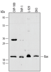
- Detection of Human/Mouse Bax by Western Blot. Western blot shows lysates of CHP-100 human neuroblastoma cell line, THP-1 human acute monocytic leukemia cell line, L-929 mouse fibroblast cell line, and DA3 mouse myeloma cell line. PVDF membrane was probed with 0.3 µg/mL of Rabbit Anti-Human/Mouse Bax Antigen Affinity-purified Polyclonal Antibody (Catalog # AF820) followed by HRP-conjugated Anti-Rabbit IgG Secondary Antibody (Catalog # HAF008). A specific band was detected for Bax at approximately 21 kDa (as indicated). This experiment was conducted under reducing conditions and using Immunoblot Buffer Group 2.

- Submitted by
- R&D Systems (provider)
- Main image
- Experimental details
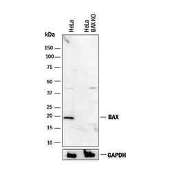
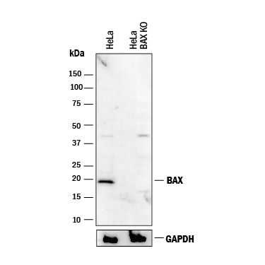
- Western Blot Shows Human Bax Specificity by Using Knockout Cell Line. Western blot shows lysates of HeLa human cervical epithelial carcinoma parental cell line and Bax knockout HeLa cell line (KO). PVDF membrane was probed with 0.5 µg/mL of Rabbit Anti-Human/Mouse Bax Antigen Affinity-purified Polyclonal Antibody (Catalog # AF820) followed by HRP-conjugated Anti-Rabbit IgG Secondary Antibody (Catalog # HAF008). A specific band was detected for Bax at approximately 20 kDa (as indicated) in the parental HeLa cell line, but is not detectable in knockout HeLa cell line. GAPDH (Catalog # AF5718) is shown as a loading control. This experiment was conducted under reducing conditions and using Immunoblot Buffer Group 1.
Supportive validation
- Submitted by
- R&D Systems (provider)
- Main image
- Experimental details
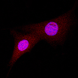
- Bax in A549 Human Cell Line. Bax was detected in immersion fixed A549 human lung carcinoma cell line using Rabbit Anti-Human/Mouse Bax Antigen Affinity-purified Polyclonal Antibody (Catalog # AF820) at 1.7 µg/mL for 3 hours at room temperature. Cells were stained using the NorthernLights™ 557-conjugated Anti-Rabbit IgG Secondary Antibody (red; Catalog # NL004) and counterstained with DAPI (blue). Specific staining was localized to cytoplasm and nuclei. View our protocol for Fluorescent ICC Staining of Cells on Coverslips.

Supportive validation
- Submitted by
- R&D Systems (provider)
- Main image

- Experimental details
- Bax in Human Breast. Bax was detected in immersion fixed paraffin-embedded sections of human breast using 15 µg/mL Rabbit Anti-Human/Mouse Bax Antigen Affinity-purified Polyclonal Antibody (Catalog # AF820) overnight at 4 °C. Tissue was stained (red) and counter-stained with hematoxylin (blue). View our protocol for Chromogenic IHC Staining of Paraffin-embedded Tissue Sections.

- Submitted by
- R&D Systems (provider)
- Main image
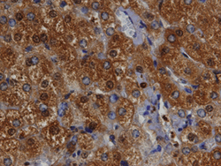
- Experimental details
- Bax in Human Liver. Bax was detected in immersion fixed paraffin-embedded sections of human liver using 10 µg/mL Rabbit Anti-Human/Mouse Bax Antigen Affinity-purified Polyclonal Antibody (Catalog # AF820) overnight at 4 °C. Tissue was stained with the Anti-Rabbit HRP-DAB Cell & Tissue Staining Kit (brown; Catalog # CTS005) and counterstained with hematoxylin (blue). View our protocol for Chromogenic IHC Staining of Paraffin-embedded Tissue Sections.
