Explore
 Validate
 Learn
Learn Western blot
Antibody data
- Antibody Data
- Antigen structure
- References [0]
- Comments [0]
- Validations
- Western blot [3]
- Immunocytochemistry [1]
- Immunohistochemistry [5]
- Flow cytometry [1]
Submit
Validation data
Reference
Comment
Report error
- Product number
- PA5-95252 - Provider product page

- Provider
- Invitrogen Antibodies
- Product name
- CtBP1 Polyclonal Antibody
- Antibody type
- Polyclonal
- Antigen
- Synthetic peptide
- Description
- Reconstitute with 0.2 mL of distilled water to yield a concentration of 500 µg/mL.
- Reactivity
- Human, Mouse, Rat
- Host
- Rabbit
- Isotype
- IgG
- Vial size
- 100 µg
- Concentration
- 500 µg/mL
- Storage
- Store at 4°C short term. For long term storage, store at -20°C, avoiding freeze/thaw cycles.
No comments: Submit comment
Supportive validation
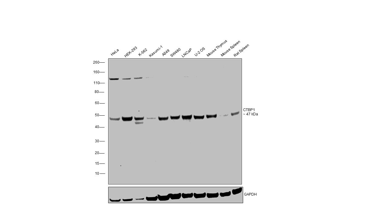
- Submitted by
- Invitrogen Antibodies (provider)
- Main image

- Experimental details
- Western blot analysis of CtBP1 in Lane 1: rat lung tissue lysate Lane 2: rat kidney tissue lysate Lane 3: COLO320 whole cell lysate Lane 4: MCF-7 whole cell lysate After Electrophoresis, proteins were transferred to a Nitrocellulose membrane at 150mA for 50-90 minutes. Electrophoresis was performed with 5-20% SDS-PAGE gel (70V, Stacking gel; 90V Resolving gel, Time: 2-3 hours), transferred to a nitrocellulose membrane and blocked using 5% Non-fat Milk/TBS (1.5 hrs at room temperature). Samples were incubated with CtBP1 polyclonal antibody (Product # PA5-95252) using a 0.5 µg/mL dilution, followed by a goat anti-rabbit IgG-HRP at a dilution of 1:10,000, and developed with enhanced chemiluminescence (ECL).
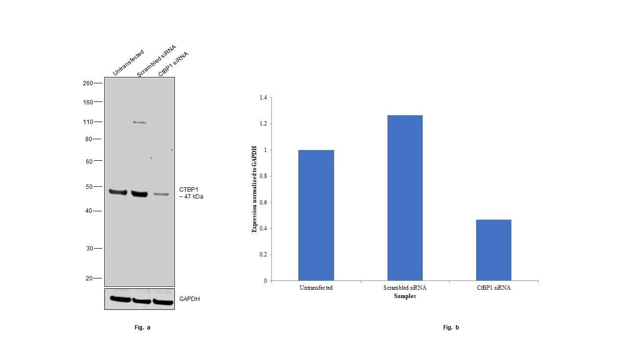
- Submitted by
- Invitrogen Antibodies (provider)
- Main image

- Experimental details
- Knockdown of CtBP1 was achieved by transfecting HEK-293 with CtBP1 specific siRNAs (Silencer® select Product # S3698, S3699). Western blot analysis (Fig. a) was performed using whole cell extracts from the CtBP1 knockdown cells (lane 3), non-targeting scrambled siRNA transfected cells (lane 2) and untransfected cells (lane 1). The blot was probed with CtBP1 Polyclonal Antibody (Product # PA5-95252, 1:2000) and Goat anti-Rabbit IgG (H+L) Superclonal™ Recombinant Secondary Antibody, HRP (Product # A27036, 1:20,000). Densitometric analysis of this western blot is shown in histogram (Fig. b). Decrease in signal upon siRNA mediated knock down confirms that antibody is specific to CtBP1.
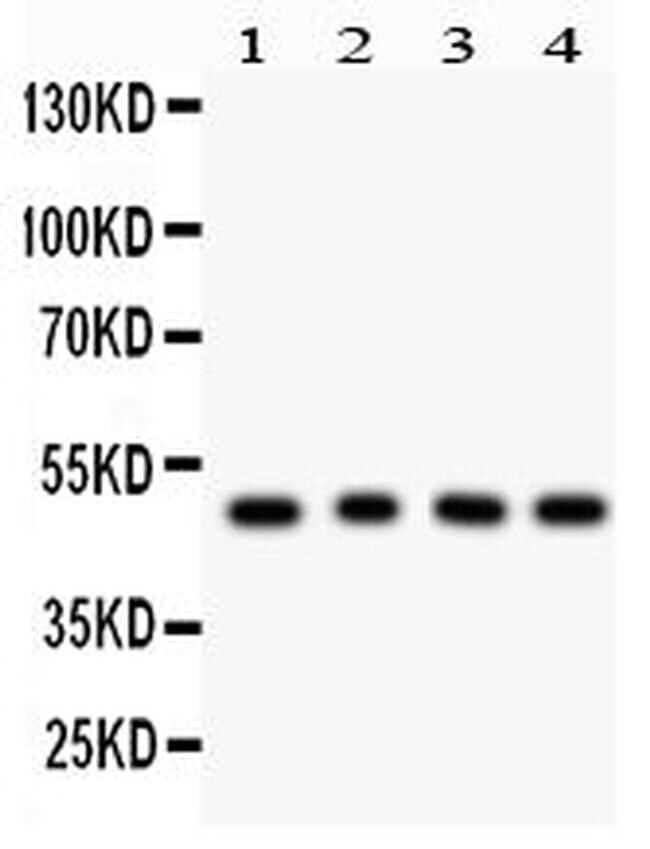
- Submitted by
- Invitrogen Antibodies (provider)
- Main image

- Experimental details
- Western blot was performed using CtBP1 Polyclonal Antibody (Product # PA5-95252) and a ~47 kDa band corresponding to CtBP1 was observed across human cell lines and mouse and rat tissues. Whole cell extracts (30 µg lysate) of HeLa (Lane 1), HEK-293 (Lane 2), K-562 (Lane 3), Kasumi-1 (Lane 4), A549 (Lane 5), SW480 (Lane 6), LNCaP (Lane 7), U-2 OS (Lane 8), Mouse Thymus (Lane 9), Mouse Spleen (Lane 10), Rat Spleen (Lane 11) were electrophoresed using NuPAGE™ 4-12% Bis-Tris Protein Gel (Product # NP0322BOX), 12 well. Resolved proteins were then transferred onto a nitrocellulose membrane (Product # IB23001) by iBlot® 2 Dry Blotting System (Product # IB21001). The blot was probed with the primary antibody (1:1000) and detected by chemiluminescence with Goat anti-Rabbit IgG (H+L) Superclonal™ Recombinant Secondary Antibody, HRP (Product # A27036, 1:20,000) using the iBright™ FL1500 Imaging System (Product # A44115). Chemiluminescent detection was performed using SuperSignal™ West Pico PLUS Chemiluminescent Substrate (Product # 34580).
Supportive validation
- Submitted by
- Invitrogen Antibodies (provider)
- Main image

- Experimental details
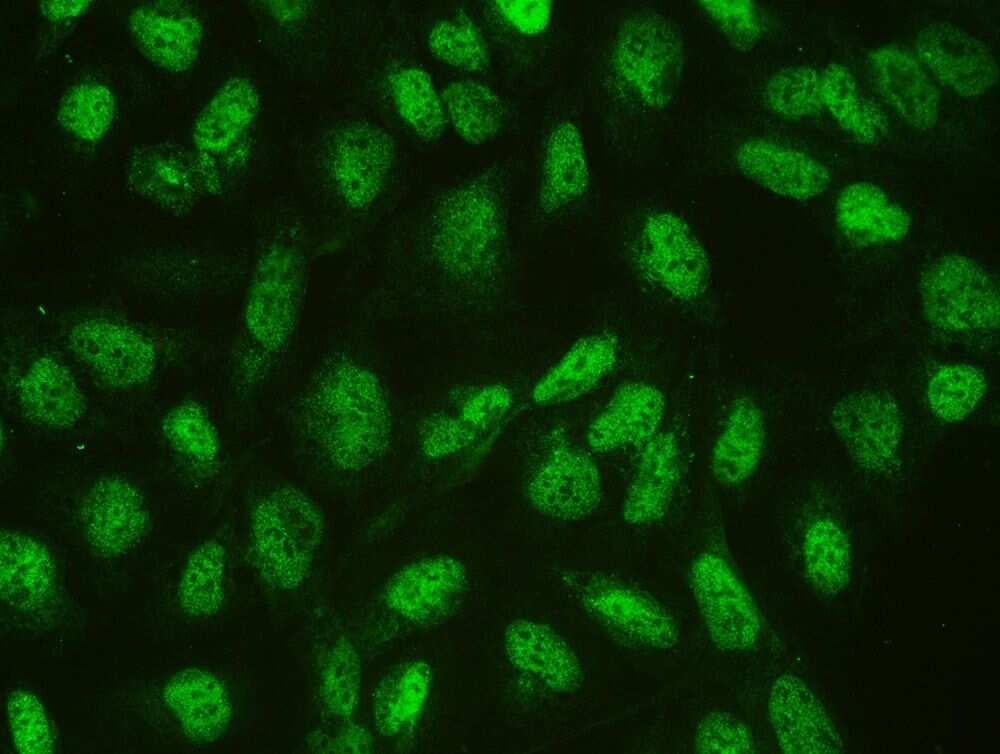
- Immunofluorescence analysis of CtBP1 in U20S cell. Antigen retrieval was performed using IHC enzyme antigen retrieval reagent and blocked with 10% goat serum. Samples were incubated with CtBP1 polyclonal antibody (Product # PA5-95252) at a 2 µg/mL dilution, followed by 488 conjugated goat anti-rabbit IgG (30 min, 37°C) using a 1:100 dilution.
Supportive validation
- Submitted by
- Invitrogen Antibodies (provider)
- Main image
- Experimental details
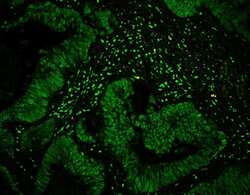
- Immunofluorescence analysis of CtBP1 in human intestinal cancer tissues. Antigen retrieval was performed on the tissue using citrate buffer (pH 6, 20 min) and blocked with 10% goat serum. Samples were incubated with CtBP1 polyclonal antibody (Product # PA5-95252) at a 1 µg/mL dilution, followed by 488 conjugated Avidin using a dilution.

- Submitted by
- Invitrogen Antibodies (provider)
- Main image
- Experimental details
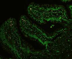
- Immunofluorescence analysis of CtBP1 in rat intestine tissues. Antigen retrieval was performed on the tissue using citrate buffer (pH 6, 20 min) and blocked with 10% goat serum. Samples were incubated with CtBP1 polyclonal antibody (Product # PA5-95252) at a 1 µg/mL dilution, followed by 488 conjugated Avidin using a dilution.

- Submitted by
- Invitrogen Antibodies (provider)
- Main image
- Experimental details
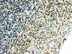
- Immunohistochemistry analysis of CtBP1 in paraffin-embedded mouse thymus tissue. Antigen retrieval was performed on the tissue using citrate buffer (pH 6, 20 min) and blocked with 10% goat serum. Samples were incubated with CtBP1 polyclonal antibody (Product # PA5-95252) at a 1 µg/mL dilution, followed by biotinylated goat anti-rabbit IgG (30 min, 37°C), and developed with Strepavidin-Biotin-Complex and DAB.
- Submitted by
- Invitrogen Antibodies (provider)
- Main image
- Experimental details
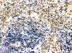
- Immunohistochemistry analysis of CtBP1 in paraffin-embedded rat spleen tissue. Antigen retrieval was performed on the tissue using citrate buffer (pH 6, 20 min) and blocked with 10% goat serum. Samples were incubated with CtBP1 polyclonal antibody (Product # PA5-95252) at a 1 µg/mL dilution, followed by biotinylated goat anti-rabbit IgG (30 min, 37°C), and developed with Strepavidin-Biotin-Complex and DAB.

- Submitted by
- Invitrogen Antibodies (provider)
- Main image
- Experimental details
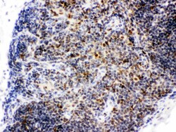
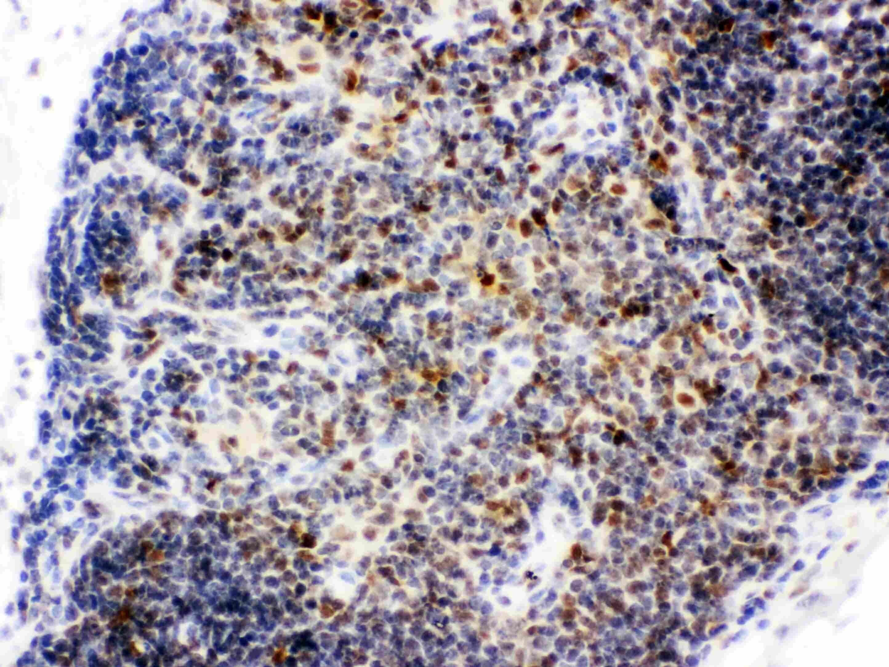
- Immunohistochemistry analysis of CtBP1 in paraffin-embedded human tonsil tissue. Antigen retrieval was performed on the tissue using citrate buffer (pH 6, 20 min) and blocked with 10% goat serum. Samples were incubated with CtBP1 polyclonal antibody (Product # PA5-95252) at a 1 µg/mL dilution, followed by biotinylated goat anti-rabbit IgG (30 min, 37°C), and developed with Strepavidin-Biotin-Complex and DAB.

Supportive validation
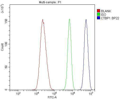
- Submitted by
- Invitrogen Antibodies (provider)
- Main image
- Experimental details
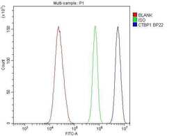
- Flow cytometry of CtBP1 in U20S cells (blue line), isotype control rabbit IgG (green line) and unlabeled (red line). Samples were blocked with 10% goat serum, incubated with CtBP1 polyclonal antibody (Product # PA5-95252) at a dilution of 1 µg (per 1x10^6 cells), followed by 488 conjugated goat anti-rabbit IgG (30 min at 20°C) using a 5-10 µg (per 1x10^6 cells) dilution.