 Explore
 Validate
 Learn
LearnCF500468
antibody from Invitrogen Antibodies
Targeting: MAPK1
ERK, ERK2, MAPK2, p41mapk, PRKM1, PRKM2
 Western blot
 Immunoprecipitation
Antibody data
- Antibody Data
- Antigen structure
- References [0]
- Comments [0]
- Validations
- Western blot [4]
- Immunohistochemistry [2]
- Flow cytometry [1]
- Other assay [1]
Submit
Validation data
Reference
Comment
Report error
- Product number
- CF500468 - Provider product page

- Provider
- Invitrogen Antibodies
- Product name
- MAPK1 Monoclonal Antibody (OTI6F8), TrueMAB™
- Antibody type
- Monoclonal
- Antigen
- Recombinant full-length protein
- Reactivity
- Human, Rat, Canine
- Host
- Mouse
- Isotype
- IgG
- Antibody clone number
- OTI6F8
- Vial size
- 100 µg
- Concentration
- 1 mg/mL
- Storage
- -20° C, Avoid Freeze/Thaw Cycles
No comments: Submit comment
Supportive validation
- Submitted by
- Invitrogen Antibodies (provider)
- Main image
- Experimental details
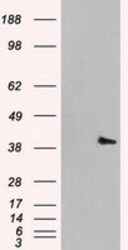
- HEK293T cells were transfected with the pCMV6-ENTRY control (Left lane) or pCMV6-ENTRY MAPK1 (RC204703, Right lane) cDNA for 48 hrs and lysed. Equivalent amounts of cell lysates (5 µg per lane) were separated by SDS-PAGE and immunoblotted with anti-MAPK1. Positive lysates LY408481 (100 µg) and LC408481 (20 µg) can be purchased separately from OriGene.

- Submitted by
- Invitrogen Antibodies (provider)
- Main image

- Experimental details
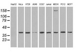
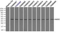
- Western blot analysis of extracts (35 µg) from 9 different cell lines by using anti-MAPK1 monoclonal antibody.
- Submitted by
- Invitrogen Antibodies (provider)
- Main image

- Experimental details
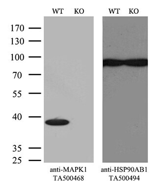
- Equivalent amounts of cell lysates (10 µg per lane) of wild-type Hela cells (WT, Cat# LC810HELA) and MAPK1-Knockout Hela cells (KO, Cat# LC810185) were separated by SDS-PAGE and immunoblotted with anti-MAPK1 monoclonal antibody TA500468. Then the blotted membrane was stripped and reprobed with anti-HSP90AB1 antibody (TA500494) as a loading control. (1:500)
- Submitted by
- Invitrogen Antibodies (provider)
- Main image

- Experimental details
- Western blot analysis of extracts (10 µg) from a mouse cell line and 3 different mouse tissues by using anti-MAPK1 monoclonal antibody. (1:200)

Supportive validation
- Submitted by
- Invitrogen Antibodies (provider)
- Main image
- Experimental details
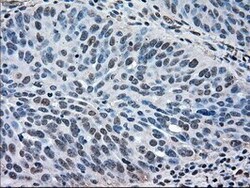
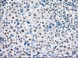
- Immunohistochemical staining of paraffin-embedded Adenocarcinoma of Human ovary tissue using anti-MAPK1 mouse monoclonal antibody. (Heat-induced epitope retrieval by 10mM citric buffer, pH6.0, 100°C for 10min, TA500468)

- Submitted by
- Invitrogen Antibodies (provider)
- Main image

- Experimental details
- Immunohistochemical staining of paraffin-embedded Adenocarcinoma of Human breast tissue using anti-MAPK1 mouse monoclonal antibody. (Heat-induced epitope retrieval by 10mM citric buffer, pH6.0, 100°C for 10min, TA500468)
Supportive validation
- Submitted by
- Invitrogen Antibodies (provider)
- Main image
- Experimental details
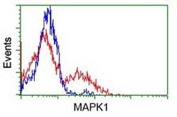
- HEK293T cells transfected with either RC204703 overexpress plasmid (Red) or empty vector control plasmid (Blue) were immunostained by anti-MAPK1 antibody (TA500468), and then analyzed by flow cytometry.

Supportive validation
- Submitted by
- Invitrogen Antibodies (provider)
- Main image
- Experimental details
- Immunoprecipitation(IP) of MAPK1 by using TrueMab monoclonal anti-MAPK1 antibodies (Negative control: IP without adding anti-MAPK1 antibody.). For each experiment, 500ul of tagged MAPK1 overexpression lysates (at 1:5 dilution with HEK293T lysate), 2 µg of anti-MAPK1 antibody and 20ul (0.1mg) of goat anti-mouse conj µgated magnetic beads were mixed and incubated overnight. After extensive wash to remove any non-specific binding, the immuno-precipitated products were analyzed with rabbit anti-DDK polyclonal antibody.
