Explore
 Validate
 Learn
Learn Western blot
Antibody data
- Antibody Data
- Antigen structure
- References [2]
- Comments [0]
- Validations
- Western blot [1]
- Immunohistochemistry [1]
Submit
Validation data
Reference
Comment
Report error
- Product number
- sc-5309 - Provider product page

- Provider
- Santa Cruz Biotechnology
- Proper citation
- Santa Cruz Biotechnology Cat#sc-5309, RRID:AB_628453
- Product name
- Anti-XRCC6
- Antibody type
- Monoclonal
- Antigen
- Recombinant full-length protein
- Reactivity
- Human
- Host
- Mouse
Submitted references Bax-inhibiting peptide protects cells from polyglutamine toxicity caused by Ku70 acetylation
Ku heterodimer binds to both ends of the Werner protein and functional interaction occurs at the Werner N-terminus.
Y Li, T Yokota, V Gama, T Yoshida, J A Gomez, K Ishikawa, H Sasaguri, H Y Cohen, D A Sinclair, H Mizusawa, S Matsuyama
Cell Death and Differentiation 2007 Sep;14(12):2058-2067
Cell Death and Differentiation 2007 Sep;14(12):2058-2067
Ku heterodimer binds to both ends of the Werner protein and functional interaction occurs at the Werner N-terminus.
Karmakar P, Snowden CM, Ramsden DA, Bohr VA
Nucleic acids research 2002 Aug 15;30(16):3583-91
Nucleic acids research 2002 Aug 15;30(16):3583-91
No comments: Submit comment
Supportive validation
- Submitted by
- per
- Main image
- Experimental details
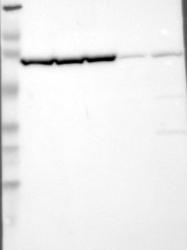
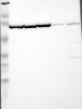
- Western blot analysis of antibody specificity using a routine panel composed of IgG/HSA-depleted human plasma and protein lysates from selected human tissues and cell lines.
- Validation comment
- Band of predicted size in kDa (+/-20%) with additional bands present.
- Primary Ab dilution
- 1:500
- Secondary Ab dilution
- 1:7000
- Lane 1
- Marker [kDa]: 230, 110, 82, 49.3, 32.2, 25.5, 17.6
- Lane 2
- RT-4
- Lane 3
- U-251MG sp
- Lane 4
- A-431
- Lane 5
- Liver
- Lane 6
- Tonsil
- Theoretical target weight
- [kDa] 70
Supportive validation
- Submitted by
- per
- Main image
- Experimental details
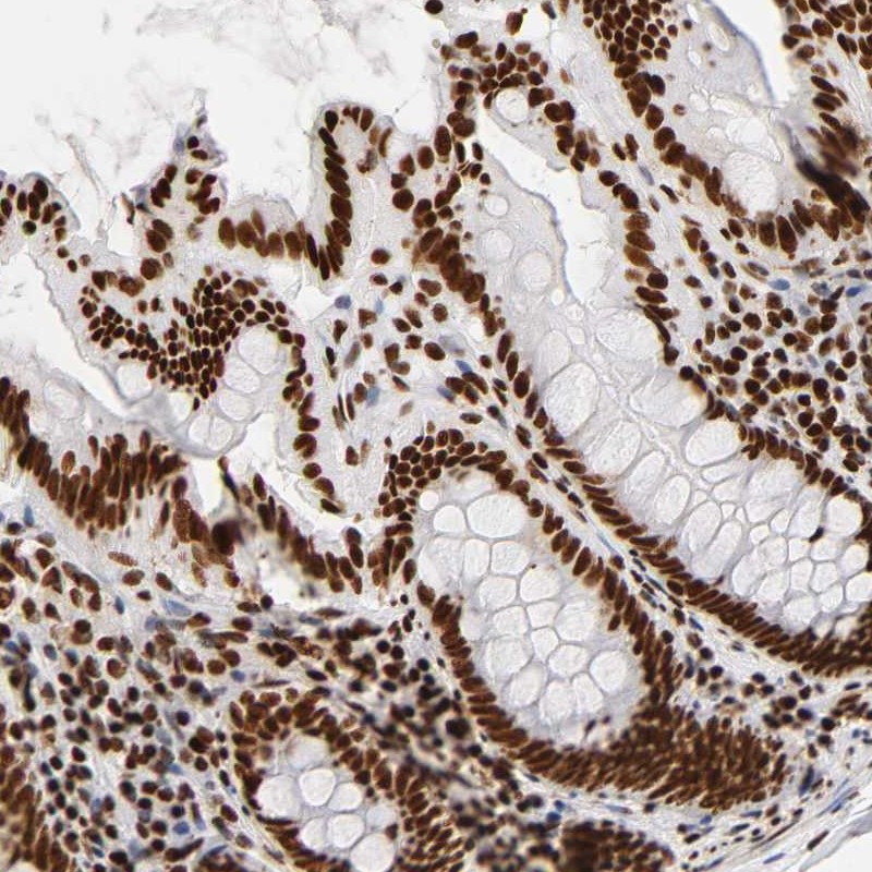
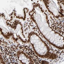
- Immunohistochemical staining of human colon shows strong nuclear positivity in glandular cells.
- Validation comment
- Two independent antibodies targeting one protein yielding similar staining patterns. Staining pattern consistent with experimental and/or bioinformatic data.