Explore
 Validate
 Learn
Learn Western blot
Antibody data
- Antibody Data
- Antigen structure
- References [2]
- Comments [0]
- Validations
- Western blot [1]
- Immunocytochemistry [1]
- Immunoprecipitation [2]
- Immunohistochemistry [1]
- Flow cytometry [3]
Submit
Validation data
Reference
Comment
Report error
- Product number
- NB100-1915 - Provider product page

- Provider
- Novus Biologicals
- Proper citation
- Novus Cat#NB100-1915, RRID:AB_10002228
- Product name
- Mouse Monoclonal Ku70/XRCC6 Antibody
- Antibody type
- Monoclonal
- Description
- Protein A purified.
- Reactivity
- Human, Mouse, Rat, Hamster, Simian, Xenopus
- Host
- Mouse
- Antigen sequence
Amino acids 506 - 541.- Isotype
- IgG
- Vial size
- 500uL
- Concentration
- 0.2 mg/ml
- Storage
- Store at 4C. Do not freeze.
Submitted references Phosphorothioate-modified CpG oligodeoxynucleotides mimic autoantigens and reveal a potential role for Toll-like receptor 9 in receptor revision.
Immortalized myogenic cells from congenital muscular dystrophy type1A patients recapitulate aberrant caspase activation in pathogenesis: a new tool for MDC1A research.
Doster A, Ziegler S, Foermer S, Rieker RJ, Heeg K, Bekeredjian-Ding I
Immunology 2013 Jun;139(2):166-78
Immunology 2013 Jun;139(2):166-78
Immortalized myogenic cells from congenital muscular dystrophy type1A patients recapitulate aberrant caspase activation in pathogenesis: a new tool for MDC1A research.
Yoon S, Stadler G, Beermann ML, Schmidt EV, Windelborn JA, Schneiderat P, Wright WE, Miller JB
Skeletal muscle 2013 Dec 6;3(1):28
Skeletal muscle 2013 Dec 6;3(1):28
No comments: Submit comment
Supportive validation
- Submitted by
- Novus Biologicals (provider)
- Main image
- Experimental details
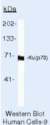
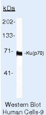
- Western Blot: Ku70/XRCC6 Antibody (N3H10) [NB100-1915] - Analysis of 50ug of HepG2 whole cell lysate per well onto a SDS-PAGE gel.
Supportive validation
- Submitted by
- Novus Biologicals (provider)
- Main image
- Experimental details
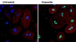
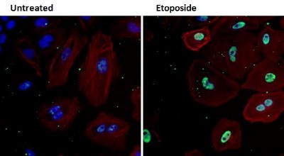
- Immunocytochemistry/Immunofluorescence: Ku70/XRCC6 Antibody (N3H10) [NB100-1915] - Analysis of Ku (green) in HeLa cells either left untreated (left panel) or treated with 50uM etoposide (right panel) for 3 hours.
Supportive validation
- Submitted by
- Novus Biologicals (provider)
- Main image
- Experimental details
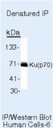
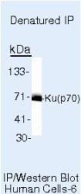
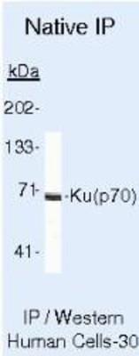
- Immunoprecipitation: Ku70/XRCC6 Antibody (N3H10) [NB100-1915] - Analysis of denatured IP.
- Submitted by
- Novus Biologicals (provider)
- Main image
- Experimental details
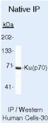
- Immunoprecipitation: Ku70/XRCC6 Antibody (N3H10) [NB100-1915] - Analysis of Ku (P70) on Native Human T47D Cells.
Supportive validation
- Submitted by
- Novus Biologicals (provider)
- Main image
- Experimental details
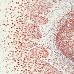
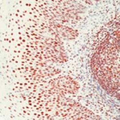
- Immunohistochemistry-Paraffin: Ku70/XRCC6 Antibody (N3H10) [NB100-1915] - Human tonsil stained with Ku antibody using peroxidase-conjugate and AEC chromogen. Note nuclear staining of epithelial cells and lymphocytes.
Supportive validation
- Submitted by
- Novus Biologicals (provider)
- Main image

- Experimental details
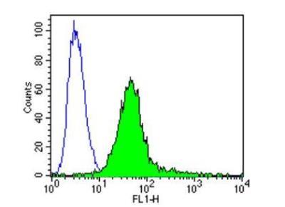
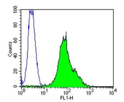
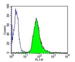
- Flow Cytometry: Ku70/XRCC6 Antibody (N3H10) [NB100-1915] - Ku (p70) in HepG2 cells compared to an isotype control (blue).
- Submitted by
- Novus Biologicals (provider)
- Main image
- Experimental details
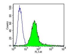
- Flow Cytometry: Ku70/XRCC6 Antibody (N3H10) [NB100-1915] - Ku (p70) in Hela cells compared to an isotype control (blue).

- Submitted by
- Novus Biologicals (provider)
- Main image
- Experimental details
- Flow Cytometry: Ku70/XRCC6 Antibody (N3H10) [NB100-1915] - Analysis of Ku (p70) in C2C12 cells compared to an isotype control (blue).