 Explore
 Validate
 Learn
LearnPA5-79579
antibody from Invitrogen Antibodies
Targeting: LAMC2
BM600-100kDa, EBR2, EBR2A, kalinin-105kDa, LAMB2T, LAMNB2, nicein-100kDa
 Western blot
Antibody data
- Antibody Data
- Antigen structure
- References [0]
- Comments [0]
- Validations
- Western blot [2]
- Immunocytochemistry [1]
- Immunohistochemistry [2]
Submit
Validation data
Reference
Comment
Report error
- Product number
- PA5-79579 - Provider product page

- Provider
- Invitrogen Antibodies
- Product name
- Laminin gamma-1,2,3 Polyclonal Antibody
- Antibody type
- Polyclonal
- Antigen
- Synthetic peptide
- Description
- This antibody reacts will all 3 isoforms of Laminin gamma: Laminin gamma-1, gamma-2 and gamma-3.
- Concentration
- 500 µg/mL
No comments: Submit comment
Supportive validation
- Submitted by
- Invitrogen Antibodies (provider)
- Main image

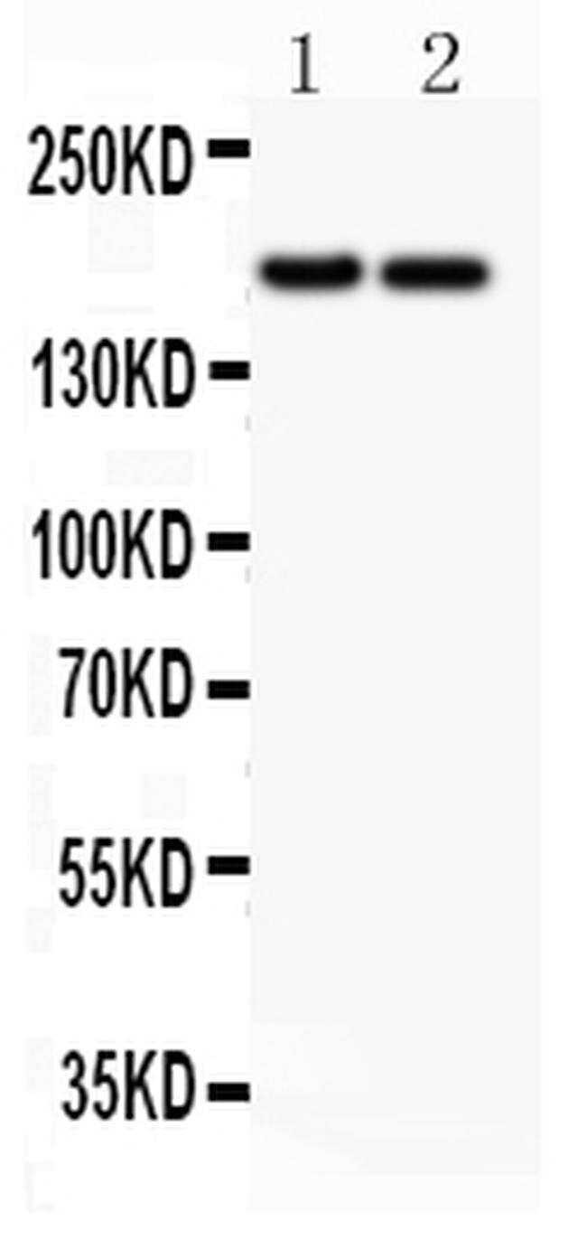
- Experimental details
- Western blot analysis of Laminin in Lane 1: HeLa whole cell lysate, Lane 2: HEPG2 whole cell lysate using 40-50 µg per well. Sample was incubated with Laminin (Product # PA5-79579) at a dilution of 0.5 µg/mL.
- Submitted by
- Invitrogen Antibodies (provider)
- Main image

- Experimental details
- Western blot was performed using anti-Laminin gamma-1,2,3 Polyclonal Antibody (Product # PA5-79579) and a 210 kDa band corresponding to Laminin gamma-1,2,3 was observed across the cell lines and tissues tested. Whole cell extracts (1% SDS) (30 µg lysate) of Hep G2 (Lane 1), HUVEC (Lane 2), A-431 (Lane 3), K-562 (Lane 4), PC-3 (Lane 5), Mouse Liver (Lane 6), Rat Liver (Lane 7), Mouse Lung (Lane 8), Rat Lung (Lane 9) were electrophoresed using NuPAGE™ 3-8% Tris-Acetate Protein Gels (Product # EA0375BOX). Resolved proteins were then transferred onto a nitrocellulose membrane (Product # IB23001) by iBlot® 2 Dry Blotting System (Product # IB21001). The blot was probed with the primary antibody (0.5 µg/mL) and detected by chemiluminescence with Goat anti-Rabbit IgG (H+L), Superclonal™ Recombinant Secondary Antibody, HRP (Product # A27036, 0.25 µg/mL, 1:10000 dilution) using the iBright FL 1000 (Product # A32752). Chemiluminescent detection was performed using Novex® ECL Chemiluminescent Substrate Reagent Kit (Product # WP20005).

Supportive validation
- Submitted by
- Invitrogen Antibodies (provider)
- Main image

- Experimental details
- Immunofluorescence analysis of Laminin gamma-1,2,3 was performed using 70% confluent log phase Hep G2 cells. The cells were fixed with 4% paraformaldehyde for 10 minutes, permeabilized with 0.1% Triton™ X-100 for 15 minutes and blocked with 2% BSA for 1 hour at room temperature. The cells were labeled with Laminin gamma-1,2,3 Polyclonal Antibody (Product # PA5-79580) at 1:100 dilution in 0.1% BSA, incubated at 4 degree celsius overnight and then with Goat anti-Rabbit IgG (H+L), Superclonal™ Recombinant Secondary Antibody, Alexa Fluor 488 conjugate (Product # A27034) at a dilution of 1:2000 for 45 minutes at room temperature (Panel a: green). Nuclei (Panel b: blue) were stained with SlowFade® Gold Antifade Mountant with DAPI (Product # S36938). F-actin (Panel c: red) was stained with Rhodamine Phalloidin (Product # R415, 1:300). Panel d represents the merged image showing cytoplasmic localization. Panel e represents K-562 cells showing no expression of Laminin gamma-1,2,3. Panel f shows control Hep G2 cells with no primary antibody to assess background. The images were captured at 60X magnification.

Supportive validation
- Submitted by
- Invitrogen Antibodies (provider)
- Main image

- Experimental details
- Immunohistochemistry analysis of Laminin on paraffin-embedded human lung cancer tissue. Sample was incubated with Laminin polyclonal antibody (Product# PA5-79579).

- Submitted by
- Invitrogen Antibodies (provider)
- Main image

- Experimental details
- Immunohistochemical analysis of Laminin in frozen section of human placenta tissues. The tissue section was blocked with 10% goat serum. The tissue section was then incubated with 1μg/mL rabbit anti-Laminin antibody (Product # PA5-79579) overnight at 4°C. Biotinylated goat anti-rabbit IgG was used as secondary antibody and incubated for 30 minutes at 37°C. The tissue section was developed using Strepavidin-Biotin-Complex (SABC) with DAB as the chromogen.
