Antibody data
- Antibody Data
- Antigen structure
- References [26]
- Comments [0]
- Validations
- Western blot [3]
- Immunocytochemistry [6]
- Flow cytometry [1]
- Other assay [15]
Submit
Validation data
Reference
Comment
Report error
- Product number
- PA3-821A - Provider product page

- Provider
- Invitrogen Antibodies
- Product name
- PPAR gamma Polyclonal Antibody
- Antibody type
- Polyclonal
- Antigen
- Synthetic peptide
- Description
- PA3-821A detects peroxisome proliferator activated receptor (PPAR) gamma from human and mouse samples. This antibody is a reproduction of PA3-821, using the same immunizing peptide and new host rabbits. PA3-821A has been successfully used in IF and Western blot procedures. Previous batches of this antibody have been used in gel shift, immunocytochemistry, and immunofluoresence procedures. By Western blot, this antibody detects a 57 kDa protein representing PPAR gamma 2 from differentiated mouse 3T3-L1 cell lysate. Immunolocalization of previous batches gives predominant nuclear staining. The PA3-821A immunizing peptide corresponds to amino acid residues 284-298 from mouse PPAR gamma 2. This sequence is completely conserved in PPAR gamma 1, but exhibits no significant homology with PPAR alpha or NUC1. This sequence is completely conserved in mouse, human, and rat.
- Reactivity
- Human, Mouse, Rat
- Host
- Rabbit
- Isotype
- IgG
- Vial size
- 100 µL
- Concentration
- Conc. Not Determined
- Storage
- -20° C, Avoid Freeze/Thaw Cycles
Submitted references Cheonggukjang-Specific Component 1,3-Diphenyl-2-Propanone as a Novel PPARα/γ Dual Agonist: An In Vitro and In Silico Study.
SetD7 (Set7/9) is a novel target of PPARγ that promotes the adaptive pancreatic β-cell glycemic response.
Decreased Substrate Stiffness Promotes a Hypofibrotic Phenotype in Cardiac Fibroblasts.
Breast cancer-associated skeletal muscle mitochondrial dysfunction and lipid accumulation is reversed by PPARG.
Effects of Pioglitazone on Glucose-Dependent Insulinotropic Polypeptide-Mediated Insulin Secretion and Adipocyte Receptor Expression in Patients With Type 2 Diabetes.
Rosiglitazone augments antioxidant response in the human trophoblast and prevents apoptosis†.
Peroxisome Proliferator-Activated Receptors (PPARs) levels in spermatozoa of normozoospermic and asthenozoospermic men.
Human Breast Cancer Xenograft Model Implicates Peroxisome Proliferator-activated Receptor Signaling as Driver of Cancer-induced Muscle Fatigue.
PPARγ regulates meibocyte differentiation and lipid synthesis of cultured human meibomian gland epithelial cells (hMGEC).
Duration of simulated microgravity affects the differentiation of mesenchymal stem cells.
Heterogeneous Differentiation of Human Mesenchymal Stem Cells in 3D Extracellular Matrix Composites.
PPAR-δ is repressed in Huntington's disease, is required for normal neuronal function and can be targeted therapeutically.
Thyroid hormone receptor sumoylation is required for preadipocyte differentiation and proliferation.
Pioglitazone-induced reductions in atherosclerosis occur via smooth muscle cell-specific interaction with PPAR{gamma}.
Activation of PPAR-γ and PTEN cascade participates in lovastatin-mediated accelerated differentiation of oligodendrocyte progenitor cells.
Ligand activation of peroxisome proliferator-activated receptor-beta/delta and inhibition of cyclooxygenase-2 enhances inhibition of skin tumorigenesis.
Effects of lysophosphatidic acid on the in vitro proliferation and differentiation of a novel porcine preadipocyte cell line.
Urine acidification has no effect on peroxisome proliferator-activated receptor (PPAR) signaling or epidermal growth factor (EGF) expression in rat urinary bladder urothelium.
Peroxisomal proliferator-activated receptor-gamma agonists induce partial reversion of epithelial-mesenchymal transition in anaplastic thyroid cancer cells.
Reciprocal expression of peroxisome proliferator-activated receptor-gamma and cyclooxygenase-2 in human term parturition.
Dexamethasone induced preadipocyte recruitment and expression of CCAAT/enhancing binding protein alpha and peroxisome proliferator activated receptor-gamma proteins in porcine stromal-vascular (S-V) cell cultures obtained before and after the onset of fetal adipogenesis.
Dexamethasone induced preadipocyte recruitment and expression of CCAAT/enhancing binding protein alpha and peroxisome proliferator activated receptor-gamma proteins in porcine stromal-vascular (S-V) cell cultures obtained before and after the onset of fetal adipogenesis.
Enhanced growth inhibition by combination differentiation therapy with ligands of peroxisome proliferator-activated receptor-gamma and inhibitors of histone deacetylase in adenocarcinoma of the lung.
Human translocation liposarcoma-CCAAT/enhancer binding protein (C/EBP) homologous protein (TLS-CHOP) oncoprotein prevents adipocyte differentiation by directly interfering with C/EBPbeta function.
MCF-7 and T47D human breast cancer cells contain a functional peroxisomal response.
MCF-7 and T47D human breast cancer cells contain a functional peroxisomal response.
Arulkumar R, Jung HJ, Noh SG, Park D, Chung HY
International journal of molecular sciences 2021 Oct 8;22(19)
International journal of molecular sciences 2021 Oct 8;22(19)
SetD7 (Set7/9) is a novel target of PPARγ that promotes the adaptive pancreatic β-cell glycemic response.
Jetton TL, Flores-Bringas P, Leahy JL, Gupta D
The Journal of biological chemistry 2021 Nov;297(5):101250
The Journal of biological chemistry 2021 Nov;297(5):101250
Decreased Substrate Stiffness Promotes a Hypofibrotic Phenotype in Cardiac Fibroblasts.
Childers RC, Lucchesi PA, Gooch KJ
International journal of molecular sciences 2021 Jun 9;22(12)
International journal of molecular sciences 2021 Jun 9;22(12)
Breast cancer-associated skeletal muscle mitochondrial dysfunction and lipid accumulation is reversed by PPARG.
Wilson HE, Stanton DA, Rellick S, Geldenhuys W, Pistilli EE
American journal of physiology. Cell physiology 2021 Apr 1;320(4):C577-C590
American journal of physiology. Cell physiology 2021 Apr 1;320(4):C577-C590
Effects of Pioglitazone on Glucose-Dependent Insulinotropic Polypeptide-Mediated Insulin Secretion and Adipocyte Receptor Expression in Patients With Type 2 Diabetes.
Tharp WG, Gupta D, Sideleva O, Deacon CF, Holst JJ, Elahi D, Pratley RE
Diabetes 2020 Feb;69(2):146-157
Diabetes 2020 Feb;69(2):146-157
Rosiglitazone augments antioxidant response in the human trophoblast and prevents apoptosis†.
Kohan-Ghadr HR, Kilburn BA, Kadam L, Johnson E, Kolb BL, Rodriguez-Kovacs J, Hertz M, Armant DR, Drewlo S
Biology of reproduction 2019 Feb 1;100(2):479-494
Biology of reproduction 2019 Feb 1;100(2):479-494
Peroxisome Proliferator-Activated Receptors (PPARs) levels in spermatozoa of normozoospermic and asthenozoospermic men.
Mousavi MS, Shahverdi A, Drevet J, Akbarinejad V, Esmaeili V, Sayahpour FA, Topraggaleh TR, Rahimizadeh P, Alizadeh A
Systems biology in reproductive medicine 2019 Dec;65(6):409-419
Systems biology in reproductive medicine 2019 Dec;65(6):409-419
Human Breast Cancer Xenograft Model Implicates Peroxisome Proliferator-activated Receptor Signaling as Driver of Cancer-induced Muscle Fatigue.
Wilson HE, Rhodes KK, Rodriguez D, Chahal I, Stanton DA, Bohlen J, Davis M, Infante AM, Hazard-Jenkins H, Klinke DJ, Pugacheva EN, Pistilli EE
Clinical cancer research : an official journal of the American Association for Cancer Research 2019 Apr 1;25(7):2336-2347
Clinical cancer research : an official journal of the American Association for Cancer Research 2019 Apr 1;25(7):2336-2347
PPARγ regulates meibocyte differentiation and lipid synthesis of cultured human meibomian gland epithelial cells (hMGEC).
Kim SW, Xie Y, Nguyen PQ, Bui VT, Huynh K, Kang JS, Brown DJ, Jester JV
The ocular surface 2018 Oct;16(4):463-469
The ocular surface 2018 Oct;16(4):463-469
Duration of simulated microgravity affects the differentiation of mesenchymal stem cells.
Xue L, Li Y, Chen J
Molecular medicine reports 2017 May;15(5):3011-3018
Molecular medicine reports 2017 May;15(5):3011-3018
Heterogeneous Differentiation of Human Mesenchymal Stem Cells in 3D Extracellular Matrix Composites.
Jung JP, Bache-Wiig MK, Provenzano PP, Ogle BM
BioResearch open access 2016;5(1):37-48
BioResearch open access 2016;5(1):37-48
PPAR-δ is repressed in Huntington's disease, is required for normal neuronal function and can be targeted therapeutically.
Dickey AS, Pineda VV, Tsunemi T, Liu PP, Miranda HC, Gilmore-Hall SK, Lomas N, Sampat KR, Buttgereit A, Torres MJ, Flores AL, Arreola M, Arbez N, Akimov SS, Gaasterland T, Lazarowski ER, Ross CA, Yeo GW, Sopher BL, Magnuson GK, Pinkerton AB, Masliah E, La Spada AR
Nature medicine 2016 Jan;22(1):37-45
Nature medicine 2016 Jan;22(1):37-45
Thyroid hormone receptor sumoylation is required for preadipocyte differentiation and proliferation.
Liu YY, Ayers S, Milanesi A, Teng X, Rabi S, Akiba Y, Brent GA
The Journal of biological chemistry 2015 Mar 20;290(12):7402-15
The Journal of biological chemistry 2015 Mar 20;290(12):7402-15
Pioglitazone-induced reductions in atherosclerosis occur via smooth muscle cell-specific interaction with PPAR{gamma}.
Subramanian V, Golledge J, Ijaz T, Bruemmer D, Daugherty A
Circulation research 2010 Oct 15;107(8):953-8
Circulation research 2010 Oct 15;107(8):953-8
Activation of PPAR-γ and PTEN cascade participates in lovastatin-mediated accelerated differentiation of oligodendrocyte progenitor cells.
Paintlia AS, Paintlia MK, Singh AK, Orak JK, Singh I
Glia 2010 Nov 1;58(14):1669-85
Glia 2010 Nov 1;58(14):1669-85
Ligand activation of peroxisome proliferator-activated receptor-beta/delta and inhibition of cyclooxygenase-2 enhances inhibition of skin tumorigenesis.
Bility MT, Zhu B, Kang BH, Gonzalez FJ, Peters JM
Toxicological sciences : an official journal of the Society of Toxicology 2010 Jan;113(1):27-36
Toxicological sciences : an official journal of the Society of Toxicology 2010 Jan;113(1):27-36
Effects of lysophosphatidic acid on the in vitro proliferation and differentiation of a novel porcine preadipocyte cell line.
Nobusue H, Kondo D, Yamamoto M, Kano K
Comparative biochemistry and physiology. Part B, Biochemistry & molecular biology 2010 Dec;157(4):401-7
Comparative biochemistry and physiology. Part B, Biochemistry & molecular biology 2010 Dec;157(4):401-7
Urine acidification has no effect on peroxisome proliferator-activated receptor (PPAR) signaling or epidermal growth factor (EGF) expression in rat urinary bladder urothelium.
Achanzar WE, Moyer CF, Marthaler LT, Gullo R, Chen SJ, French MH, Watson LM, Rhodes JW, Kozlosky JC, White MR, Foster WR, Burgun JJ, Car BD, Cosma GN, Dominick MA
Toxicology and applied pharmacology 2007 Sep 15;223(3):246-56
Toxicology and applied pharmacology 2007 Sep 15;223(3):246-56
Peroxisomal proliferator-activated receptor-gamma agonists induce partial reversion of epithelial-mesenchymal transition in anaplastic thyroid cancer cells.
Aiello A, Pandini G, Frasca F, Conte E, Murabito A, Sacco A, Genua M, Vigneri R, Belfiore A
Endocrinology 2006 Sep;147(9):4463-75
Endocrinology 2006 Sep;147(9):4463-75
Reciprocal expression of peroxisome proliferator-activated receptor-gamma and cyclooxygenase-2 in human term parturition.
Dunn-Albanese LR, Ackerman WE 4th, Xie Y, Iams JD, Kniss DA
American journal of obstetrics and gynecology 2004 Mar;190(3):809-16
American journal of obstetrics and gynecology 2004 Mar;190(3):809-16
Dexamethasone induced preadipocyte recruitment and expression of CCAAT/enhancing binding protein alpha and peroxisome proliferator activated receptor-gamma proteins in porcine stromal-vascular (S-V) cell cultures obtained before and after the onset of fetal adipogenesis.
Hausman GJ
General and comparative endocrinology 2003 Aug;133(1):61-70
General and comparative endocrinology 2003 Aug;133(1):61-70
Enhanced growth inhibition by combination differentiation therapy with ligands of peroxisome proliferator-activated receptor-gamma and inhibitors of histone deacetylase in adenocarcinoma of the lung.
Chang TH, Szabo E
Clinical cancer research : an official journal of the American Association for Cancer Research 2002 Apr;8(4):1206-12
Clinical cancer research : an official journal of the American Association for Cancer Research 2002 Apr;8(4):1206-12
Human translocation liposarcoma-CCAAT/enhancer binding protein (C/EBP) homologous protein (TLS-CHOP) oncoprotein prevents adipocyte differentiation by directly interfering with C/EBPbeta function.
Adelmant G, Gilbert JD, Freytag SO
The Journal of biological chemistry 1998 Jun 19;273(25):15574-81
The Journal of biological chemistry 1998 Jun 19;273(25):15574-81
MCF-7 and T47D human breast cancer cells contain a functional peroxisomal response.
Kilgore MW, Tate PL, Rai S, Sengoku E, Price TM
Molecular and cellular endocrinology 1997 May 16;129(2):229-35
Molecular and cellular endocrinology 1997 May 16;129(2):229-35
MCF-7 and T47D human breast cancer cells contain a functional peroxisomal response.
Kilgore MW, Tate PL, Rai S, Sengoku E, Price TM
Molecular and cellular endocrinology 1997 May 16;129(2):229-35
Molecular and cellular endocrinology 1997 May 16;129(2):229-35
No comments: Submit comment
Supportive validation
- Submitted by
- Invitrogen Antibodies (provider)
- Main image
- Experimental details
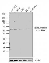
- Western blot analysis was performed on whole cell extracts (20 µg lysate) of NIH/3T3 (Lane 1), HeLa (Lane 2), K562 (Lane 3), PC-12 (lane 4) and U-87 MG (lane 5). The blots were probed with Anti-PPAR gamma Rabbit Polyclonal Antibody (Product # PA3-821A, 1:500-1:1500 dilution) and detected by chemiluminescence using Goat anti-Rabbit IgG (H+L) Superclonal™ Secondary Antibody, HRP conjugate (Product # A27036, 0.4 µg/mL, 1:2500 dilution). A 54 kDa band corresponding to PPAR gamma was observed across cell lines tested. Known quantity of protein samples were electrophoresed using Novex® NuPAGE® 4-12 % Bis-Tris gel (Product # NP0322BOX), XCell SureLock™ Electrophoresis System (Product # EI0002) and Novex® Sharp Pre-Stained Protein Standard (Product # LC5800). Resolved proteins were then transferred onto a nitrocellulose membrane with iBlot® 2 Dry Blotting System (Product # IB21001). The membrane was probed with the relevant primary and secondary Antibody following blocking with 5 % skimmed milk. Chemiluminescent detection was performed using Pierce™ ECL Western Blotting Substrate (Product # 32106).
- Submitted by
- Invitrogen Antibodies (provider)
- Main image

- Experimental details
- Western blot analysis of PPAR gamma-2 was performed by loading 20 µg of indicated whole cell lysate and 5 µL of PageRuler Plus Prestained Protein Ladder (Product # 26619) per well onto a 4-20% Tris-Glycine polyacrylamide gel (Product # WT4202BX10). Proteins were transferred to a nitrocellulose membrane using the G2 Blotter (Product # 62288), and blocked with 5% BSA in TBST for 1 hour at room temperature. PPAR gamma was detected at ~57 kDa using a PPAR Gamma rabbit polyclonal antibody (Product # PA3-821A) at a dilution of 1:500 in blocking buffer overnight at 4C on a rocking platform, followed by a Goat anti-Rabbit IgG (H+L) Superclonal Secondary Antibody, HRP conjugate (Product # A27036) at a dilution of 1:1000 for at least 30 minutes at room temperature. Chemiluminescent detection was performed using SuperSignal West Pico (Product # 34078).
- Submitted by
- Invitrogen Antibodies (provider)
- Main image

- Experimental details
- Western blot analysis of PPAR gamma-2 was performed by loading 20 µg of indicated whole cell lysate and 5 µL of PageRuler Plus Prestained Protein Ladder (Product # 26619) per well onto a 4-20% Tris-Glycine polyacrylamide gel (Product # WT4202BX10). Proteins were transferred to a nitrocellulose membrane using the G2 Blotter (Product # 62288), and blocked with 5% BSA in TBST for 1 hour at room temperature. PPAR gamma was detected at ~57 kDa using a PPAR Gamma rabbit polyclonal antibody (Product # PA3-821A) at a dilution of 1:500 in blocking buffer overnight at 4C on a rocking platform, followed by a Goat anti-Rabbit IgG (H+L) Superclonal™ Secondary Antibody, HRP conjugate (Product # A27036) at a dilution of 1:1000 for at least 30 minutes at room temperature. Chemiluminescent detection was performed using SuperSignal West Pico (Product # 34078).
Supportive validation
- Submitted by
- Invitrogen Antibodies (provider)
- Main image

- Experimental details
- Immunofluorescent analysis of PPAR gamma (green) showing positive staining in the nucleus and cytoplasm of 3T3-L1 cells (right) compared with a negative control in the absence of primary antibody (left). Formalin-fixed cells were permeabilized with 0.1% Triton X-100 in TBS for 5-10 minutes, blocked with 3% BSA-PBS for 30 minutes at room temperature and probed with a PPAR gamma polyclonal antibody (Product # PA3-821A) in 3% BSA-PBS at a dilution of 1:200 and incubated overnight at 4 ºC in a humidified chamber. Cells were washed with PBST and incubated with a DyLight 488-conjugated goat-anti-rabbit IgG secondary antibody in PBS at room temperature in the dark. F-actin (red) was stained with a fluorescent red phalloidin and nuclei (blue) were stained with DAPI for 5-10 minutes in the dark. Images were taken at a magnification of 60x.
- Submitted by
- Invitrogen Antibodies (provider)
- Main image
- Experimental details
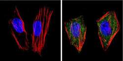
- Immunofluorescent analysis of PPAR gamma (green) showing positive staining in the nucleus and cytoplasm of Hela cells (right) compared with a negative control in the absence of primary antibody (left). Formalin-fixed cells were permeabilized with 0.1% Triton X-100 in TBS for 5-10 minutes, blocked with 3% BSA-PBS for 30 minutes at room temperature and probed with a PPAR gamma polyclonal antibody (Product # PA3-821A) in 3% BSA-PBS at a dilution of 1:200 and incubated overnight at 4 ºC in a humidified chamber. Cells were washed with PBST and incubated with a DyLight 488-conjugated goat-anti-rabbit IgG secondary antibody in PBS at room temperature in the dark. F-actin (red) was stained with a fluorescent red phalloidin and nuclei (blue) were stained with DAPI for 5-10 minutes in the dark. Images were taken at a magnification of 60x.
- Submitted by
- Invitrogen Antibodies (provider)
- Main image
- Experimental details
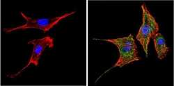
- Immunofluorescent analysis of PPAR gamma (green) showing positive staining in the nucleus and cytoplasm of NIH-3T3 cells (right) compared with a negative control in the absence of primary antibody (left). Formalin-fixed cells were permeabilized with 0.1% Triton X-100 in TBS for 5-10 minutes, blocked with 3% BSA-PBS for 30 minutes at room temperature and probed with a PPAR gamma polyclonal antibody (Product # PA3-821A) in 3% BSA-PBS at a dilution of 1:200 and incubated overnight at 4 ºC in a humidified chamber. Cells were washed with PBST and incubated with a DyLight 488-conjugated goat-anti-rabbit IgG secondary antibody in PBS at room temperature in the dark. F-actin (red) was stained with a fluorescent red phalloidin and nuclei (blue) were stained with DAPI for 5-10 minutes in the dark. Images were taken at a magnification of 60x.
- Submitted by
- Invitrogen Antibodies (provider)
- Main image

- Experimental details
- Immunofluorescence analysis of PPAR Gamma was done on 70% confluent log phase HeLa cells. The cells were fixed with 4% paraformaldehyde for 15 minutes, permeabilized with 0.25% Triton™ X-100 for 10 minutes, and blocked with 5% BSA for 1 hour at room temperature. The cells were labeled with PPAR gamma Rabbit Polyclonal Antibody (Product # PA3-821A) at 1:250 dilution in 0.1% BSA and incubated for 3 hours at room temperature and then labeled with Goat anti-Rabbit IgG (H+L) Superclonal™ Secondary Antibody, Alexa Fluor® 488 conjugate (Product # A27034) at a dilution of 1:2000 for 45 minutes at room temperature (Panel a: green). Nuclei (Panel b: blue) were stained with SlowFade® Gold Antifade Mountant with DAPI (Product # S36938). F-actin (Panel c: red) was stained with Rhodamine Phalloidin (Product # R415, 1:300). Panel d is a merged image showing Nuclear localization. Panel e is a no primary antibody control. The images were captured at 60X magnification.
- Submitted by
- Invitrogen Antibodies (provider)
- Main image
- Experimental details
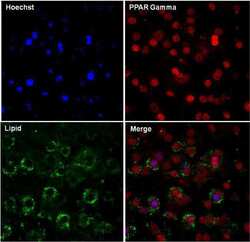
- Immunofluorescent analysis of PPAR gamma-2 (red) in 3T3-L1 differentiated day 7 cells. The cells were fixed with 4% paraformaldehyde in PBS for 15 minutes at room temperature, permeabilized with 0.1% Triton X-100 for 15 minutes, and blocked with 3% BSA for 30 minutes at room temperature. Cells were stained with a PPAR gamma-2 rabbit polyclonal antibody (Product # PA3-821A) at a dilution of 1:200 in blocking buffer for 1 hour at room temperature, and then incubated with a Goat anti-Rabbit IgG (H+L) Secondary Antibody, Dylight 680 (Product # 35568) at a dilution of 1:1000 for at least 30 minutes at room temperature in the dark (red). Lipids (green) were stained with HCS LipidTOX neutral lipid stain (Product # H34475) at a dilution of 1:200 for at least 30 minutes at room temperature in the dark. Nuclei (blue) were stained with Hoechst 33342 (Product # 62249). Images were taken on a EVOS FL Auto Imaging System at 20X magnification.
- Submitted by
- Invitrogen Antibodies (provider)
- Main image

- Experimental details
- Immunofluorescent analysis of PPAR gamma-2 (green) in 3T3-L1 Undifferentiated (left panel) and Differentiated day 7 cells (right panel. The cells were fixed with 4% paraformaldehyde in PBS for 15 minutes at room temperature, permeabilized with 0.1% Triton X-100 for 15 minutes, and blocked with 3% BSA for 30 minutes at room temperature. Cells were stained with a PPAR gamma rabbit polyclonal antibody (Product # PA3-821A) at a dilution of 1:200 in blocking buffer for 1 hour at room temperature, and then incubated with a Goat anti-Rabbit IgG (H+L) Superclonal Secondary Antibody, Alexa Fluor® 488 conjugate (Product # A27034) at a dilution of 1:1000 for at least 30 minutes at room temperature in the dark. Nuclei (blue) were stained with Hoechst 33342 (Product # 62249). Images were taken on a EVOS FL Auto Imaging System at 60X magnification.
Supportive validation
- Submitted by
- Invitrogen Antibodies (provider)
- Main image
- Experimental details
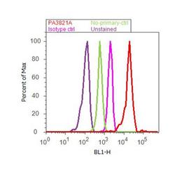
- Flow cytometry analysis of PPAR gamma was done on Jurkat cells. Cells were fixed with 70% ethanol for 10 minutes, permeabilized with 0.25% Triton™ X-100 for 20 minutes, and blocked with 5% BSA for 30 minutes at room temperature. Cells were labeled with PPAR gamma Rabbit Polyclonal Antibody (PA3821A, red histogram) or with rabbit isotype control (pink histogram) at 3-5 ug/million cells in 2.5% BSA. After incubation at room temperature for 2 hours, the cells were labeled with Alexa Fluor® 488 Goat Anti-Rabbit Secondary Antibody (A11008) at a dilution of 1:400 for 30 minutes at room temperature. The representative 10,000 cells were acquired and analyzed for each sample using an Attune® Acoustic Focusing Cytometer. The purple histogram represents unstained control cells and the green histogram represents no-primary-antibody control.
Supportive validation
- Submitted by
- Invitrogen Antibodies (provider)
- Main image

- Experimental details
- NULL
- Submitted by
- Invitrogen Antibodies (provider)
- Main image
- Experimental details
- NULL
- Submitted by
- Invitrogen Antibodies (provider)
- Main image
- Experimental details
- NULL
- Submitted by
- Invitrogen Antibodies (provider)
- Main image
- Experimental details
- NULL
- Submitted by
- Invitrogen Antibodies (provider)
- Main image
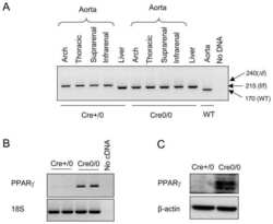
- Experimental details
- NULL
- Submitted by
- Invitrogen Antibodies (provider)
- Main image

- Experimental details
- NULL
- Submitted by
- Invitrogen Antibodies (provider)
- Main image

- Experimental details
- NULL
- Submitted by
- Invitrogen Antibodies (provider)
- Main image
- Experimental details
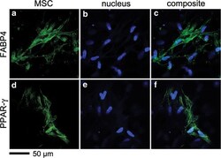
- FIG. 4. Expression of FABP4 and PPAR-gamma proteins in 3D ColI composites after 28 days of culture. Immunofluorescence was visualized by MPLSM. FABP4 (a-c) and PPAR-gamma (d-f) . Adipogenic protein markers (green) and nuclei (blue); scale bar 50 mum.
- Submitted by
- Invitrogen Antibodies (provider)
- Main image
- Experimental details
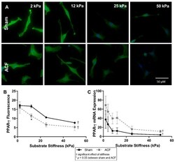
- Figure 7 ( A ) PPAR-gamma fluorescence (green) with DAPI nuclear counterstain (blue) and quantification ( B ) show a decrease in intensity with increasing stiffness. ( C ) mRNA expression is also inversely related with substrate stiffness. The highest expression and fluorescence and mRNA levels appears on the softest substrates. * indicates p < 0.05 when comparing sham vs. ACF. + indicates a significant effect of substrate stiffness.
- Submitted by
- Invitrogen Antibodies (provider)
- Main image

- Experimental details
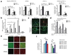
- Figure 8 DPP increased transcriptional activity of PPARalpha and PPARgamma. For luciferase, the 3x PPRE-TK-LUC plasmid and PPARalpha ( A ) or PPARgamma ( B ) expression vector were transfected into Ac2F cells. Twenty-four hours after the transfection, the cells were treated with DPP or agonists (WY14643 10 uM and Rosiglitazone 10 uM, respectively) for 5 h. Values are expressed as the mean +- SEM of two independent replications. ( C ) The nuclear levels of PPARalpha and PPARgamma in Ac2F cells were analysed by western blot and band intensity were quantified by ImageJ program. Transcription factor II B (TFIIB) was used as loading control. Statistical results of one-factor ANOVA followed by the Bonferroni test: ** p
- Submitted by
- Invitrogen Antibodies (provider)
- Main image
- Experimental details
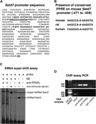
- Figure 1 Nuclear binding assays. A , a partial SetD7 mouse promoter sequence curated using NM_080,793 as DNA reference sequence. The SetD7 PPRE sequence is bolded and underlined . The DNA sequences used for the primers designed for the ChIP assay PCR are bolded and italicized . B , consensus PPRE sequence on the SetD7 promoter: A putative PPARgamma response element was found using MatInspector software on the SetD7 promoter region that was conserved between mouse, rat, and human SetD7 promoter sequences. C , EMSA and super-shift assay of SetD7 PPRE: PAGE purified infrared dye (IRD-700) tagged oligos for SetD7 PPRE were annealed and EMSA was performed using nuclear extracts prepared from INS-1 cells as detailed in the Experimental procedures section. For the super-shift assay, INS-1 cell nuclear extracts were preincubated with a PPARgamma specific antibody or mouse nonimmune serum on ice for 30 min before adding infrared dye (IRD-700) tagged oligos for SetD7 PPRE. D , upper panel , ChIP assay PCR showing 144 base pairs (bp) SetD7 PPRE positive band: 300-400 bp chromatin preparations of betaTC6 cells were prepared as described in the Experimental procedures section. The prepared chromatin samples were immunoprecipitated using anti-PPARgamma versus a control nonimmune IgG, followed by PCR of the immunoprecipitated and nonimmunoprecipitated DNA (input DNA) using flanking primer pairs to the mouse SetD7-PPRE. The shown representative bands are the 144 bp (expected length) PCR prod
- Submitted by
- Invitrogen Antibodies (provider)
- Main image
- Experimental details
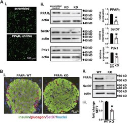
- Figure 2 SetD7 is reduced in PPARgamma KD and PANC PPARgamma KO beta-cells. A , i , 10x overview of live INS-1 cells expressing GFP-tagged PPARgamma shRNA or scrambled (Scr) shRNA. A , ii , representative WB and band quantitation for PPARgamma, SetD7 and Pdx1 relative to the scrambled control, * p < 0.001. B , i , Representative immunofluorescence fields, imaged were under identical conditions, from 8-week-old WT and PANC PPARgamma KO islets stained for insulin ( green ), SetD7 ( magenta ), and glucagon ( red ). B , ii , representative WB showing protein levels of PPARgamma and SetD7 in the islets of 8-week-old WT and PANC PPARgamma KO mice. B , iii , quantitative band analysis of SetD7 in WT and PANC PPARgamma KO mice. * p < 0.001.
- Submitted by
- Invitrogen Antibodies (provider)
- Main image
- Experimental details
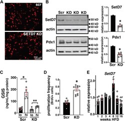
- Figure 5 SetD7 expression in beta-cells of sham and 60% Px in rats post 14-days surgery. A , SetD7 immunohistochemistry: Pancreatic sections of sham and 60% Px rats were stained for SetD7 (indirect immunoperoxidase staining). Representative pancreatic islet fields demonstrate enhanced SetD7 staining in the 60% Px islets compared with sham controls correlating with beta-cell functional compensation. B , SetD7 immunofluorescence analysis: Representative pancreatic islet fields from sham control and 60% Px rats were immunolabeled for insulin ( green ), glucagon ( red ), SetD7 ( magenta ), and nuclei (DAPI, blue ) demonstrating enhanced nuclear SetD7 staining in beta-cells of 60% Px rats compared with sham controls under conditions of sustained beta-cell compensation. C , WB and band quantitation: Lysates from isolated islets from the pancreas of sham & 60% Px rats were analyzed for PPARgamma and SetD7. The data are representative of three separate WBs and demonstrate enhanced expression of PPARgamma and SetD7 in islets of Px rats compared with sham controls. * p < 0.002.
- Submitted by
- Invitrogen Antibodies (provider)
- Main image
- Experimental details
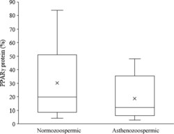
- Figure 2. PPARg protein expression in human spermatozoa based on flow cytometric analysis. Percentage of PPARg protein-positive spermatozoa in normozoospermic (n = 39) and asthenozoospermic (n = 40) groups after flow cytometric analysis (mean+-SEM). The proportion of sperm positive for PPARg protein tended to be higher in normozoospermic than in asthenozoospermic men (P = 0.066).
- Submitted by
- Invitrogen Antibodies (provider)
- Main image
- Experimental details
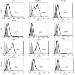
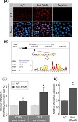
- Figure 8. Rosiglitazone effect on nuclear translocation of PPARgamma and CAT transcription in JEG-3 cells. (A) Immunofluorescence proximity ligation assay (PLA) for global transcriptional activity of PPARgamma in JEG-3 cells. Using primary antibodies against PPARgamma and RNA polymerase-2 (POL2), the technique enables visualization of molecular proximity (potentially at gene promoters) at single molecule resolution. Negative control (negative) imaged cells processed after treatment with rosiglitazone (Rosi.) without primary antibodies. (B) ChIP assays demonstrating the occupancy of PPARgamma and RNA polymerase-2 (POL2) on the promoters of the CAT gene. The DNA fragments were IP using specific antibodies against PPARgamma, POL2, or control IgG, and analyzed by qPCR. Input DNA served as an internal control. The target promoter region upstream of the transcription start site of the human CAT gene (verified by MEME SUITE; upper panel) was targeted. (C) Results were normalized to input DNA, and the enrichments were calculated relative to untreated (N/T) control. (D) RT-qPCR was performed in parallel to measure CAT mRNA expression. N = 3; * P < 0.05 vs N/T control, N/T = no-treatment, Rosi. = Rosiglitazone, scale bar = 100 mum, bars present mean +- SEM.
 Explore
 Validate
 Learn
Learn Western blot