Antibody data
- Antibody Data
- Antigen structure
- References [1]
- Comments [0]
- Validations
- Western blot [3]
- ELISA [1]
- Immunocytochemistry [1]
- Immunohistochemistry [2]
- Flow cytometry [1]
Submit
Validation data
Reference
Comment
Report error
- Product number
- MA5-31940 - Provider product page

- Provider
- Invitrogen Antibodies
- Product name
- TUBB1 Monoclonal Antibody (2A1A9)
- Antibody type
- Monoclonal
- Antigen
- Purifed from natural sources
- Description
- MA5-31940 has been tested in indirect ELISA.
- Reactivity
- Human, Mouse, Rat
- Host
- Mouse
- Isotype
- IgG
- Antibody clone number
- 2A1A9
- Vial size
- 100 µL
- Concentration
- 1 mg/mL
- Storage
- Store at 4°C short term. For long term storage, store at -20°C, avoiding freeze/thaw cycles.
Submitted references Temporal resolution of gene derepression and proteome changes upon PROTAC-mediated degradation of BCL11A protein in erythroid cells.
Mehta S, Buyanbat A, Kai Y, Karayel O, Goldman SR, Seruggia D, Zhang K, Fujiwara Y, Donovan KA, Zhu Q, Yang H, Nabet B, Gray NS, Mann M, Fischer ES, Adelman K, Orkin SH
Cell chemical biology 2022 Aug 18;29(8):1273-1287.e8
Cell chemical biology 2022 Aug 18;29(8):1273-1287.e8
No comments: Submit comment
Supportive validation
- Submitted by
- Invitrogen Antibodies (provider)
- Main image

- Experimental details
- Western blot analysis of TUBB1 in human TUBB1 recombinant protein. Sample was incubated with TUBB1 monoclonal antibody (Product # MA5-31940) using a dilution of 1:500-1:2000.
- Submitted by
- Invitrogen Antibodies (provider)
- Main image

- Experimental details
- Western blot analysis of TUBB1 in HEK293 (1) and TUBB1 cell lysate. Samples were incubated with TUBB1 monoclonal antibody (Product # MA5-31940) using a dilution of 1:500-1:2000.
- Submitted by
- Invitrogen Antibodies (provider)
- Main image

- Experimental details
- Western blot analysis of TUBB1 in K562 (1), HepG2 (2), A431 (3), Jurkat (4), HeLa (5), NIH/3T3 (6), Cos7 (7) and PC12 (8) cell lysate. Samples were incubated with TUBB1 monoclonal antibody (Product # MA5-31940) using a dilution of 1:500-1:2000.
Supportive validation
- Submitted by
- Invitrogen Antibodies (provider)
- Main image

- Experimental details
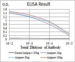
- ELISA analysis of TUBB1 in Control Antigen (black line, 100 ng); Antigen (purple line, 10 ng); Antigen (blue line, 50 ng); Antigen (red line, 100 ng). Samples were incubated with TUBB1 monoclonal antibody (Product # MA5-31940) using a dilution of 1:10,000.
Supportive validation
- Submitted by
- Invitrogen Antibodies (provider)
- Main image

- Experimental details
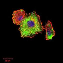
- Immunocytochemistry analysis of TUBB1 in Hela cells (green). Sample was incubated with TUBB1 monoclonal antibody (Product # MA5-31940) using a dilution of 1:200-1:1000 followed by DRAQ5 fluorescent DNA dye (blue), and Alexa Fluor- 555 phalloidin (red labeled actin filaments) .
Supportive validation
- Submitted by
- Invitrogen Antibodies (provider)
- Main image

- Experimental details
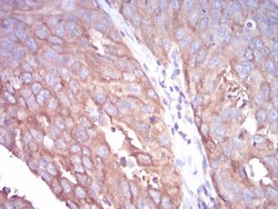
- Immunohistochemistry analysis of TUBB1 in paraffin-embedded ovarian cancer tissue. Sample was incubated with TUBB1 monoclonal antibody (Product # MA5-31940) using a dilution of 1:200-1:1000 followed by DAB staining.
- Submitted by
- Invitrogen Antibodies (provider)
- Main image

- Experimental details
- Immunohistochemistry analysis of TUBB1 in paraffin-embedded ovarian cancer tissue. Sample was incubated with TUBB1 monoclonal antibody (Product # MA5-31940) using a dilution of 1:200-1:1000 followed by DAB staining.
Supportive validation
- Submitted by
- Invitrogen Antibodies (provider)
- Main image

- Experimental details
- Flow cytometry of TUBB1 in A431 cells (green). Sample was incubated with TUBB1 monoclonal antibody (Product # MA5-31940) using a dilution of 1:200-1:400 followed by negative control (red).
 Explore
 Validate
 Learn
Learn Western blot