 Explore
 Validate
 Learn
Learn Western blot
Antibody data
- Antibody Data
- Antigen structure
- References [1]
- Comments [0]
- Validations
- Western blot [2]
- Immunohistochemistry [1]
Submit
Validation data
Reference
Comment
Report error
- Product number
- MAB3928 - Provider product page

- Provider
- Novus Biologicals
- Product name
- Mouse Monoclonal Annexin A2 Antibody
- Antibody type
- Monoclonal
- Description
- Protein A or G purified from hybridoma culture supernatant. Detects human Annexin A2 in direct ELISAs and Western blots. In direct ELISAs and Western blots, less than 25% cross-reactivity with recombinant human (rh) Annexin A1, A7, A9, A11, and A13 is observed. No cross-reactivity with rhAnnexin A3, A4, A6, A8, or A10 is observed.
- Reactivity
- Human, Mouse, Rat
- Host
- Mouse
- Isotype
- IgG
- Vial size
- 100 ug
- Concentration
- LYOPH
- Storage
- Use a manual defrost freezer and avoid repeated freeze-thaw cycles. 12 months from date of receipt, -20 to -70 degreesC as supplied. 1 month, 2 to 8 degreesC under sterile conditions after reconstitution. 6 months, -20 to -70 degreesC under sterile conditions after reconstitution.
Submitted references Annexin A3 Knockdown Suppresses Lung Adenocarcinoma.
Liu YF, Liu QQ, Zhang YH, Qiu JH
Analytical cellular pathology (Amsterdam) 2016;2016:4131403
Analytical cellular pathology (Amsterdam) 2016;2016:4131403
No comments: Submit comment
Supportive validation
- Submitted by
- Novus Biologicals (provider)
- Main image

- Experimental details
- Detection of Human, Mouse, and Rat Annexin A2 by Western Blot. Western blot shows lysates of HeLa human cervical epithelial carcinoma cell line, A431 human epithelial carcinoma cell line, C2C12 mouse myoblast cell line, and L6 rat myoblast cell line. PVDF Membrane was probed with 0.1 µg/mL of Mouse Anti-Human/Mouse/Rat Annexin A2 Monoclonal Antibody (Catalog # MAB3928) followed by HRP-conjugated Anti-Mouse IgG Secondary Antibody (Catalog # HAF007). A specific band was detected for Annexin A2 at approximately 40 kDa (as indicated). This experiment was conducted under reducing conditions and using Immunoblot Buffer Group 1.

- Submitted by
- Novus Biologicals (provider)
- Main image

- Experimental details
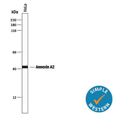
- Detection of Human Annexin A2 by Simple WesternTM. Simple Western lane view shows lysates of HeLa human cervical epithelial carcinoma cell line, loaded at 0.2 mg/mL. A specific band was detected for Annexin A2 at approximately 43 kDa (as indicated) using 1 µg/mL of Mouse Anti-Human/Mouse/Rat Annexin A2 Monoclonal Antibody (Catalog # MAB3928). This experiment was conducted under reducing conditions and using the 12-230 kDa separation system.
Supportive validation
- Submitted by
- Novus Biologicals (provider)
- Main image

- Experimental details
- Annexin A2 in Human Liver. Annexin A2 was detected in immersion fixed paraffin-embedded sections of human liver using Mouse Anti-Human/Mouse/Rat Annexin A2 Monoclonal Antibody (Catalog # MAB3928) at 15 µg/mL overnight at 4 °C. Before incubation with the primary antibody, tissue was subjected to heat-induced epitope retrieval using Antigen Retrieval Reagent-Basic (Catalog # CTS013). Tissue was stained using the Anti-Mouse HRP-DAB Cell & Tissue Staining Kit (brown; Catalog # CTS002) and counterstained with hematoxylin (blue). Specific staining was localized to plasma membranes and cytoplasm of hepatocytes. View our protocol for Chromogenic IHC Staining of Paraffin-embedded Tissue Sections.
