Explore
 Validate
 Learn
Learn Western blot
 Immunoprecipitation
Antibody data
- Antibody Data
- Antigen structure
- References [0]
- Comments [0]
- Validations
- Western blot [3]
- Immunocytochemistry [1]
- Immunohistochemistry [2]
Submit
Validation data
Reference
Comment
Report error
- Product number
- PA5-27085 - Provider product page

- Provider
- Invitrogen Antibodies
- Product name
- Annexin A2 Polyclonal Antibody
- Antibody type
- Polyclonal
- Antigen
- Synthetic peptide
- Description
- Recommended positive controls: A549, HeLa, HCT116, human ANXA2-transfected 293T. Predicted reactivity: Mouse (100%), Rat (100%), Dog (100%), Pig (100%), Sheep (100%), Bovine (100%). Store product as a concentrated solution. Centrifuge briefly prior to opening the vial.
- Reactivity
- Human
- Host
- Rabbit
- Isotype
- IgG
- Vial size
- 100 µL
- Concentration
- 0.75 mg/mL
- Storage
- Store at 4°C short term. For long term storage, store at -20°C, avoiding freeze/thaw cycles.
No comments: Submit comment
Supportive validation
- Submitted by
- Invitrogen Antibodies (provider)
- Main image
- Experimental details
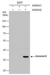
- Western Blot analysis of Annexin A2 was performed by separating 30 µg of non-transfected (-) and human Annexin I or Annexin II-transfected (+) whole cell extracts by 10% SDS-PAGE. Proteins were transferred to a membrane and probed with a Annexin A2 Polyclonal Antibody (Product # PA5-27085) at a dilution of 1:1000. The HRP-conjugated anti-rabbit IgG antibody was used to detect the primary antibody.

- Submitted by
- Invitrogen Antibodies (provider)
- Main image

- Experimental details
- Annexin A2 Polyclonal Antibody detects ANXA2 protein by western blot analysis. A. 30 µg A549 whole cell lysate/extract. B. 30 µg HCT116 whole cell lysate/extract.10% SDS-PAGE. Annexin A2 Polyclonal Antibody (Product # PA5-27085) dilution: 1:1,000. The HRP-conjugated anti-rabbit IgG antibody was used to detect the primary antibody.

- Submitted by
- Invitrogen Antibodies (provider)
- Main image
- Experimental details
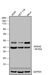
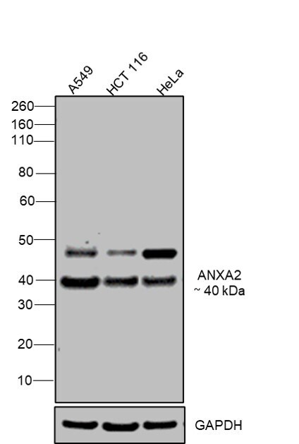
- Western blot was performed using Anti-Annexin A2 Polyclonal Antibody(Product # PA5-27085) and a 40kDa band corresponding to Annexin A2 was observed across cell lines tested. Whole cell extracts (30 µg lysate) of A549 (Lane 1), HCT 116 (Lane 2), HeLa (Lane 3) were electrophoresed using NuPAGE™ 4-12% Bis-Tris Protein Gel (Product # NP0322BOX). Resolved proteins were then transferred onto a Nitrocellulose membrane (Product # IB23001) by iBlot® 2 Dry Blotting System (Product # IB21001). The blot was probed with the primary antibody (1:1000 Dilution) and detected by chemiluminescence with Goat anti-Rabbit IgG (H+L) Superclonal™ Recombinant Secondary Antibody, HRP (Product # A27036,1:4000 dilution) using the iBright FL 1000 (Product # A32752). Chemiluminescent detection was performed using Novex® ECL Chemiluminescent Substrate Reagent Kit (Product # WP20005).
Supportive validation
- Submitted by
- Invitrogen Antibodies (provider)
- Main image

- Experimental details
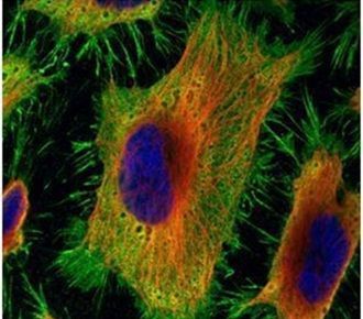
- Immunofluorescent analysis of Annexin II in paraformaldehyde-fixed HeLa cells using an Annexin II polyclonal antibody (Product # PA5-27085) (Green) at a 1:500 dilution. Alpha-tubulin filaments were labeled with Product # PA5-29281 (Red) at a 1:2000.
Supportive validation
- Submitted by
- Invitrogen Antibodies (provider)
- Main image

- Experimental details
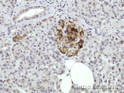
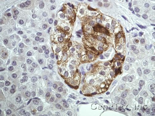
- Immunohistochemical analysis of paraffin-embedded Human pancreas tissue, using ANXA2 (Product # PA5-27085) antibody at 1:100 dilution. Antigen Retrieval: EDTA based buffer, pH 8.0, 15 min.
- Submitted by
- Invitrogen Antibodies (provider)
- Main image
- Experimental details
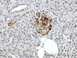
- Immunohistochemical analysis of paraffin-embedded Human pancreas tissue, using ANXA2 (Product # PA5-27085) antibody at 1:100 dilution. Antigen Retrieval: EDTA based buffer, pH 8.0, 15 min.