 Explore
 Validate
 Learn
Learn Western blot
Antibody data
- Antibody Data
- Antigen structure
- References [9]
- Comments [0]
- Validations
- Western blot [2]
- Flow cytometry [2]
- Blocking/Neutralizing [1]
Submit
Validation data
Reference
Comment
Report error
- Product number
- MAB141-100 - Provider product page

- Provider
- R&D Systems
- Product name
- Human B7-2/CD86 Antibody
- Antibody type
- Monoclonal
- Description
- Protein A or G purified from ascites. Detects human B7-2/CD86 in direct ELISAs and Western blots. In direct ELISAs, no cross-reactivity with recombinant human (rh) B7-1, recombinant mouse B7-2, recombinant rat B7-2, rhB7-H1, rhB7-H2, rhB7-H3, rhB7-H3b, rhB7-H4, or rhB7-L2 is observed.
- Reactivity
- Human
- Host
- Mouse
- Conjugate
- Unconjugated
- Antigen sequence
P42081- Isotype
- IgG
- Antibody clone number
- 37301
- Vial size
- 100 ug
- Concentration
- LYOPH
- Storage
- Use a manual defrost freezer and avoid repeated freeze-thaw cycles. 12 months from date of receipt, -20 to -70 °C as supplied. 1 month, 2 to 8 °C under sterile conditions after reconstitution. 6 months, -20 to -70 °C under sterile conditions after reconstitution.
Submitted references Comparison of the biological characteristics of human mesenchymal stem cells derived from exfoliated deciduous teeth, bone marrow, gingival tissue, and umbilical cord.
Endothelial extracellular vesicles modulate the macrophage phenotype: Potential implications in atherosclerosis.
Human trophoblast cells induced MDSCs from peripheral blood CD14(+) myelomonocytic cells via elevated levels of CCL2.
Tumor-activated TCRγδ⁺ T cells from gastric cancer patients induce the antitumor immune response of TCRαβ⁺ T cells via their antigen-presenting cell-like effects.
Endoneurial macrophages induce perineural invasion of pancreatic cancer cells by secretion of GDNF and activation of RET tyrosine kinase receptor.
ICOS-LICOS interaction is critically involved in TGN1412-mediated T-cell activation.
Phagocytosis of apoptotic cells modulates mesenchymal stem cells osteogenic differentiation to enhance IL-17 and RANKL expression on CD4+ T cells.
Critical role of lipid rafts in CD154-mediated T cell signaling.
Circulating myeloid dendritic cells of advanced cancer patients result in reduced activation and a biased cytokine profile in invariant NKT cells.
Li J, Xu SQ, Zhao YM, Yu S, Ge LH, Xu BH
Molecular medicine reports 2018 Dec;18(6):4969-4977
Molecular medicine reports 2018 Dec;18(6):4969-4977
Endothelial extracellular vesicles modulate the macrophage phenotype: Potential implications in atherosclerosis.
He S, Wu C, Xiao J, Li D, Sun Z, Li M
Scandinavian journal of immunology 2018 Apr;87(4):e12648
Scandinavian journal of immunology 2018 Apr;87(4):e12648
Human trophoblast cells induced MDSCs from peripheral blood CD14(+) myelomonocytic cells via elevated levels of CCL2.
Zhang Y, Qu D, Sun J, Zhao L, Wang Q, Shao Q, Kong B, Zhang Y, Qu X
Cellular & molecular immunology 2016 Sep;13(5):615-27
Cellular & molecular immunology 2016 Sep;13(5):615-27
Tumor-activated TCRγδ⁺ T cells from gastric cancer patients induce the antitumor immune response of TCRαβ⁺ T cells via their antigen-presenting cell-like effects.
Mao C, Mou X, Zhou Y, Yuan G, Xu C, Liu H, Zheng T, Tong J, Wang S, Chen D
Journal of immunology research 2014;2014:593562
Journal of immunology research 2014;2014:593562
Endoneurial macrophages induce perineural invasion of pancreatic cancer cells by secretion of GDNF and activation of RET tyrosine kinase receptor.
Cavel O, Shomron O, Shabtay A, Vital J, Trejo-Leider L, Weizman N, Krelin Y, Fong Y, Wong RJ, Amit M, Gil Z
Cancer research 2012 Nov 15;72(22):5733-43
Cancer research 2012 Nov 15;72(22):5733-43
ICOS-LICOS interaction is critically involved in TGN1412-mediated T-cell activation.
Weissmüller S, Semmler LY, Kalinke U, Christians S, Müller-Berghaus J, Waibler Z
Blood 2012 Jun 28;119(26):6268-77
Blood 2012 Jun 28;119(26):6268-77
Phagocytosis of apoptotic cells modulates mesenchymal stem cells osteogenic differentiation to enhance IL-17 and RANKL expression on CD4+ T cells.
Tso GH, Law HK, Tu W, Chan GC, Lau YL
Stem cells (Dayton, Ohio) 2010 May;28(5):939-54
Stem cells (Dayton, Ohio) 2010 May;28(5):939-54
Critical role of lipid rafts in CD154-mediated T cell signaling.
El Fakhry Y, Alturaihi H, Diallo D, Merhi Y, Mourad W
European journal of immunology 2010 Mar;40(3):770-9
European journal of immunology 2010 Mar;40(3):770-9
Circulating myeloid dendritic cells of advanced cancer patients result in reduced activation and a biased cytokine profile in invariant NKT cells.
van der Vliet HJ, Wang R, Yue SC, Koon HB, Balk SP, Exley MA
Journal of immunology (Baltimore, Md. : 1950) 2008 Jun 1;180(11):7287-93
Journal of immunology (Baltimore, Md. : 1950) 2008 Jun 1;180(11):7287-93
No comments: Submit comment
Supportive validation
- Submitted by
- R&D Systems (provider)
- Main image
- Experimental details
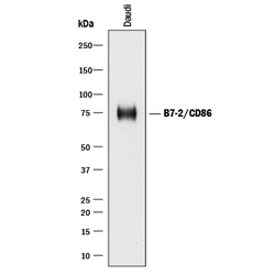
- Detection of Human B7-2/CD86 by Western Blot. Western blot shows lysates of Daudi human Burkitt's lymphoma cell line. PVDF membrane was probed with 5 µg/mL of Mouse Anti-Human B7-2/CD86 Monoclonal Antibody (Catalog # MAB141) followed by HRP-conjugated Anti-Mouse IgG Secondary Antibody (Catalog # HAF018). A specific band was detected for B7-2/CD86 at approximately 75 kDa (as indicated). This experiment was conducted under reducing conditions and using Immunoblot Buffer Group 1.
- Submitted by
- R&D Systems (provider)
- Main image
- Experimental details
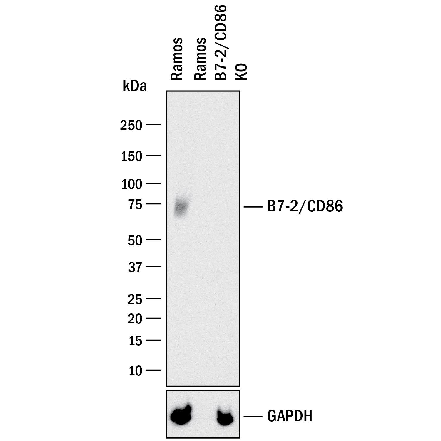
- Western Blot Shows Human B7-2/CD86 Specificity by Using Knockout Cell Line. Western blot shows lysates of Ramos human Burkitt's lymphoma parental cell line and B7-2/CD86 knockout Ramos cell line (KO). PVDF membrane was probed with 5 µg/mL of Mouse Anti-Human B7-2/CD86 Monoclonal Antibody (Catalog # MAB141) followed by HRP-conjugated Anti-Mouse IgG Secondary Antibody (Catalog # HAF018). A specific band was detected for B7-2/CD86 at approximately 74 kDa (as indicated) in the parental Ramos cell line, but is not detectable in knockout Ramos cell line. GAPDH (Catalog # AF5718) is shown as a loading control. This experiment was conducted under reducing conditions and using Immunoblot Buffer Group 1.
Supportive validation
- Submitted by
- R&D Systems (provider)
- Main image

- Experimental details
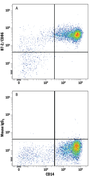
- Detection of B7-2/CD86 in Human Blood Monocytes by Flow Cytometry. Human peripheral blood monocytes were stained with Mouse Anti-Human CD14 PE-conjugated Monoclonal Antibody (Catalog # FAB3832P) and either (A) Mouse Anti-Human B7-2/CD86 Monoclonal Antibody (Catalog # MAB141) or (B) Mouse IgG1 Isotype Control (Catalog # MAB002) followed by Allophycocyanin-conjugated Anti-Mouse IgG Secondary Antibody (Catalog # F0101B).
- Submitted by
- R&D Systems (provider)
- Main image

- Experimental details
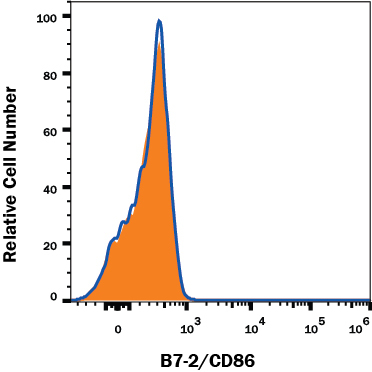
- B7-2/CD86 Specificity is Shown by Flow Cytometry in Knockout Cell Line. B7-2/CD86 knockout Ramos human lymphoma cell line was stained with Mouse Anti-Human B7-2/CD86 Monoclonal Antibody (Catalog # MAB141, filled histogram) or isotype control antibody (Catalog # MAB002, open histogram) followed by PE-conjugated Goat anti-Mouse IgG Secondary Antibody (Catalog # F0102B). No staining in the B7-2/CD86 knockout Ramos cell line was observed. View our protocol for Staining Membrane-associated Proteins.
Supportive validation
- Submitted by
- R&D Systems (provider)
- Main image

- Experimental details
- Cell IL-2 Secretion Induced by B7-2/CD86 and Neutralization by Human B7-2/CD86 Antibody. Recombinant Human B7-2/CD86 Fc Chimera induces IL-2 secretion in the Jurkat human acute T cell leukemia cell line in a dose-dependent manner (orange line), as measured by the Human IL-2 Quantikine kit (Catalog # D2050). Under these conditions, IL-2 secretion elicited by B7-2/ CD86 is neutralized (green line) by increasing concentrations of Mouse Anti-Human B7-2/CD86 Monoclonal Antibody (Catalog # MAB141). The ND50 is typically 0.5-2.5 μg/mL.
