Antibody data
- Antibody Data
- Antigen structure
- References [3]
- Comments [0]
- Validations
- Western blot [2]
- Immunocytochemistry [1]
- Immunohistochemistry [5]
- Other assay [1]
Submit
Validation data
Reference
Comment
Report error
- Product number
- UM500008 - Provider product page

- Provider
- OriGene
- Proper citation
- OriGene Cat#UM500008, RRID:AB_2629023
- Product name
- Anti-ERCC1 mouse monoclonal antibody, clone 4F9
- Antibody type
- Monoclonal
- Description
- Anti-ERCC1 mouse monoclonal antibody, clone 4F9
- Host
- Mouse
- Conjugate
- Unconjugated
- Epitope
- ERCC1
- Isotype
- IgG
- Antibody clone number
- 4F9
- Vial size
- 100 µl
- Concentration
- 0.5-1.0 mg/ml
Submitted references Measuring ERCC1 protein expression in cancer specimens: validation of a novel antibody.
ERCC1 is a prognostic biomarker in locally advanced head and neck cancer: results from a randomised, phase II trial.
Using protein microarray technology to screen anti-ERCC1 monoclonal antibodies for specificity and applications in pathology.
Smith DH, Fiehn AM, Fogh L, Christensen IJ, Hansen TP, Stenvang J, Nielsen HJ, Nielsen KV, Hasselby JP, Brünner N, Jensen SS
Scientific reports 2014 Mar 7;4:4313
Scientific reports 2014 Mar 7;4:4313
ERCC1 is a prognostic biomarker in locally advanced head and neck cancer: results from a randomised, phase II trial.
Bauman JE, Austin MC, Schmidt R, Kurland BF, Vaezi A, Hayes DN, Mendez E, Parvathaneni U, Chai X, Sampath S, Martins RG
British journal of cancer 2013 Oct 15;109(8):2096-105
British journal of cancer 2013 Oct 15;109(8):2096-105
Using protein microarray technology to screen anti-ERCC1 monoclonal antibodies for specificity and applications in pathology.
Ma D, Baruch D, Shu Y, Yuan K, Sun Z, Ma K, Hoang T, Fu W, Min L, Lan ZS, Wang F, Mull L, He WW
BMC biotechnology 2012 Nov 21;12:88
BMC biotechnology 2012 Nov 21;12:88
No comments: Submit comment
Supportive validation
- Submitted by
- OriGene (provider)
- Main image

- Experimental details
- Western blot analysis of extracts (35ug) from 9 different cell lines by using anti-ERCC1 monoclonal antibody (Clone 4F9).
- Validation comment
- WB
- Submitted by
- OriGene (provider)
- Main image

- Experimental details
- Western blot of human tissue lysates (15ug) from 10 different tissues (1: Testis, 2: Omentum, 3: Uterus, 4: Breast, 5: Brain, 6: Liver, 7: Ovary, 8: Thyroid, 9: Colon, 10: Spleen ). Diluation: 1:500.
- Validation comment
- WB
Supportive validation
- Submitted by
- OriGene (provider)
- Main image

- Experimental details
- Immunofluorescent staining of HeLa cells using anti-ERCC1 mouse monoclonal antibody (UM500008, green, 1:100). Actin filaments were labeled with Alexa Fluor? 594 Phalloidin (red), and nuclear with DAPI (blue). Scale bar, 20?m.
- Validation comment
- IF
Supportive validation
- Submitted by
- OriGene (provider)
- Main image

- Experimental details
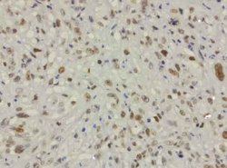
- Immunohistochemical staining of paraffin-embedded carcinoma of lung tissue (large cell) using anti-ERCC1 mouse monoclonal antibody. (Clone 4F9, dilution 1:200; heat-induced epitope retrieval by 1mM EDTA in 10mM Tris Buffer, pH 8.0, 110C for 3min)
- Submitted by
- OriGene (provider)
- Main image

- Experimental details
- Immunohistochemical staining of formalin fixed paraffin-embedded carcinoma of lung tissue (squamous cell) using anti-ERCC1 mouse monoclonal antibody. (Clone 4F9, dilution 1:50; heat-induced epitope retrieval by 1mM EDTA in 10mM Tris Buffer, pH 8.0, 110C for 3min)
- Submitted by
- OriGene (provider)
- Main image

- Experimental details
- Immunohistochemical staining of paraffin-embedded adenocarcinoma of lung (bronchioloalveolar) using anti-ERCC1 mouse monoclonal antibody. (Clone 4F9, dilution 1:50; heat-induced epitope retrieval by 1mM EDTA in 10mM Tris Buffer, pH 8.0, 110C for 3min)
- Submitted by
- OriGene (provider)
- Main image

- Experimental details
- Immunohistochemical staining of FFPE normal adjacent lung from tumor specimen using heat-induced epitope retrieval HIER at 120C for 3min with Accel buffer pH8.7, mouse monoclonal antibody anti-ERCC1 clone 4F9 was used at 1ug/mL. Strong nuclear stain in lung pneumocytes and macrophages.
- Validation comment
- IHC
- Submitted by
- OriGene (provider)
- Main image

- Experimental details
- Immunohistochemical staining of FFPE endometrial carcinoma using heat-induced epitope retrieval HIER at 120C for 3min with Accel buffer pH8.7, mouse monoclonal antibody anti-ERCC1 clone 4F9 was used at 1ug/mL. Strong nuclear stain in tumor cells.
- Validation comment
- IHC
Supportive validation
- Submitted by
- OriGene (provider)
- Main image

- Experimental details
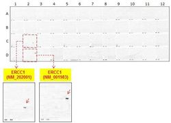
- OriGene overexpression protein microarray chip was immunostained with UltraMAB anti-ERCC1 mouse monoclonal antibody (Clone 4F9). The positive reactive proteins are highlighted with red arrows in the enlarged subarray. Other positive controls spotted in this subarray are serial dilutions of mouse IgG as controls.
- Validation comment
- 10K-CHIP
 Explore
 Validate
 Learn
Learn Western blot