Explore
 Validate
 Learn
Learn Western blot
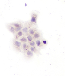
 Immunocytochemistry
Antibody data
- Antibody Data
- Antigen structure
- References [4]
- Comments [0]
- Validations
- Western blot [1]
Submit
Validation data
Reference
Comment
Report error
- Product number
- PA1036-1 - Provider product page

- Provider
- Boster Biological Technology
- Product name
- Anti-GAD67/GAD1 Antibody
- Antibody type
- Polyclonal
- Description
- Polyclonal antibody for GAD67/GAD1 detection. Host: Rabbit.Size: 100μg/vial. Tested applications: WB. Reactive species: Human. GAD67/GAD1 information: Molecular Weight: 66897 MW; Tissue Specificity: Isoform 3 is expressed in pancreatic islets, testis, adrenal cortex, and perhaps other endocrine tissues, but not in brain.
- Reactivity
- Human, Mouse, Rat
- Host
- Rabbit
- Vial size
- 100μg/vial
- Concentration
- Add 0.2ml of distilled water will yield a concentration of 500ug/ml.
- Storage
- At -20°C for one year. After reconstitution, at 4°C for one month. It can also be aliquoted and stored frozen at -20°C for a longer time. Avoid repeated freezing and thawing.
- Handling
- Add 0.2ml of distilled water will yield a concentration of 500ug/ml.
Submitted references Alteration of GABAergic signaling is associated with anxiety-like behavior in temporal lobe epilepsy mice.
Neuronal nitric oxide synthase contributes to pentylenetetrazole-kindling-induced hippocampal neurogenesis.
Integration of donor mesenchymal stem cell-derived neuron-like cells into host neural network after rat spinal cord transection.
In vitro expression of cytokeratin 18, 19 and tube formation of adipose-derived stem cells induced by the breast epithelial cell line HBL-100.
Zhu X, Yao Y, Li X, Dong J, Zhang A
Progress in neuro-psychopharmacology & biological psychiatry 2019 Jul 13;93:141-148
Progress in neuro-psychopharmacology & biological psychiatry 2019 Jul 13;93:141-148
Neuronal nitric oxide synthase contributes to pentylenetetrazole-kindling-induced hippocampal neurogenesis.
Zhu X, Dong J, Shen K, Bai Y, Chao J, Yao H
Brain research bulletin 2016 Mar;121:138-47
Brain research bulletin 2016 Mar;121:138-47
Integration of donor mesenchymal stem cell-derived neuron-like cells into host neural network after rat spinal cord transection.
Zeng X, Qiu XC, Ma YH, Duan JJ, Chen YF, Gu HY, Wang JM, Ling EA, Wu JL, Wu W, Zeng YS
Biomaterials 2015 Jun;53:184-201
Biomaterials 2015 Jun;53:184-201
In vitro expression of cytokeratin 18, 19 and tube formation of adipose-derived stem cells induced by the breast epithelial cell line HBL-100.
Yang J, Xiong L, Wang R, Yuan Q, Xia Y, Sun J, Horch RE
Journal of cellular and molecular medicine 2015 Dec;19(12):2827-31
Journal of cellular and molecular medicine 2015 Dec;19(12):2827-31
No comments: Submit comment
Supportive validation
- Submitted by
- Boster Biological Technology (provider)
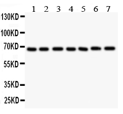
- Main image

- Experimental details
- Western blot analysis of GAD67 using anti- GAD67 antibody (PA1036-1). Electrophoresis was performed on a 5-20% SDS-PAGE gel at 70V (Stacking gel) / 90V (Resolving gel) for 2-3 hours. The sample well of each lane was loaded with 50ug of sample under reducing conditions. Lane 1: Rat Brain Tissue Lysate, Lane 2: Rat Testis Tissue Lysate, Lane 3: MCF-7 Whole Cell Lysate, Lane 4: MM231 Whole Cell Lysate, Lane 5: HELA Whole Cell Lysate, Lane 6: SMMC Whole Cell Lysate, Lane 7: COLO320 Whole Cell Lysate. After Electrophoresis, proteins were transferred to a Nitrocellulose membrane at 150mA for 50-90 minutes. Blocked the membrane with 5% Non-fat Milk/ TBS for 1.5 hour at RT. The membrane was incubated with rabbit anti- GAD67 antigen affinity purified polyclonal antibody (Catalog # PA1036-1) at 0.5 μg/mL overnight at 4°C, then washed with TBS-0.1%Tween 3 times with 5 minutes each and probed with a goat anti-rabbit IgG-HRP secondary antibody at a dilution of 1:10000 for 1.5 hour at RT. The signal is developed using an Enhanced Chemiluminescent detection (ECL) kit (Catalog # EK1002) with Tanon 5200 system. A specific band was detected for GAD67 at approximately 67KD. The expected band size for GAD67 is at 67KD.
- Additional image