 Explore
 Validate
 Learn
Learn12433-1-AP
antibody from Invitrogen Antibodies
Targeting: HOMER1
HOMER-1B, SYN47, Ves-1
 Western blot Immunocytochemistry
 Immunoprecipitation Immunohistochemistry Flow cytometry Other assay
Antibody data
- Antibody Data
- Antigen structure
- References [0]
- Comments [0]
- Validations
- Western blot [5]
- Immunocytochemistry [3]
- Immunohistochemistry [8]
- Flow cytometry [1]
- Other assay [1]
Submit
Validation data
Reference
Comment
Report error
- Product number
- 12433-1-AP - Provider product page

- Provider
- Invitrogen Antibodies
- Product name
- HOMER1 Polyclonal Antibody
- Antibody type
- Polyclonal
- Antigen
- Other
- Description
- Immunogen sequence: MGEQPIFST RAHVFQIDPN TKKNWVPTSK HAVTVSYFYD STRNVYRIIS LDGSKAIINS TITPNMTFTK TSQKFGQWAD SRANTVYGLG FSSEHHLSKF AEKFQEFKEA ARLAKEKSQE KMELTSTPSQ ESAGGDLQSP LTPESINGTD DERTPDVTQN SEPRAEPTQN ALPFSHSSAI SKHWEAELAT LKGNNAKLTA ALLESTANVK QWKQQLAAYQ EEAERLHKRV TELECVSSQA NAVHTHKTEL NQTIQELEET LKLKEEEIER LKQEIDNARE LQEQRDSLTQ KLQEVEIRNK DLEGQLSDLE QRLEKSQNEQ EAFRNNLKTL LEILDGKIFE LTELRDNLAK LLECS (1-354 aa encoded by BC015502)
- Reactivity
- Human, Mouse, Rat
- Host
- Rabbit
- Isotype
- IgG
- Vial size
- 150 µL
- Concentration
- 0.46 mg/mL
- Storage
- -20°C
No comments: Submit comment
Supportive validation
- Submitted by
- Invitrogen Antibodies (provider)
- Main image

- Experimental details
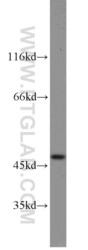
- WB result of anti-HOMER1 (12433-1-AP) with mouse brain lysate and human brain lysate.

- Submitted by
- Invitrogen Antibodies (provider)
- Main image
- Experimental details
- Human brain tissue were subjected to SDS PAGE followed by western blot with 12433-1-AP (HOMER1 antibody) at dilution of 1:1000 incubated at room temperature for 1.5 hours.

- Submitted by
- Invitrogen Antibodies (provider)
- Main image

- Experimental details
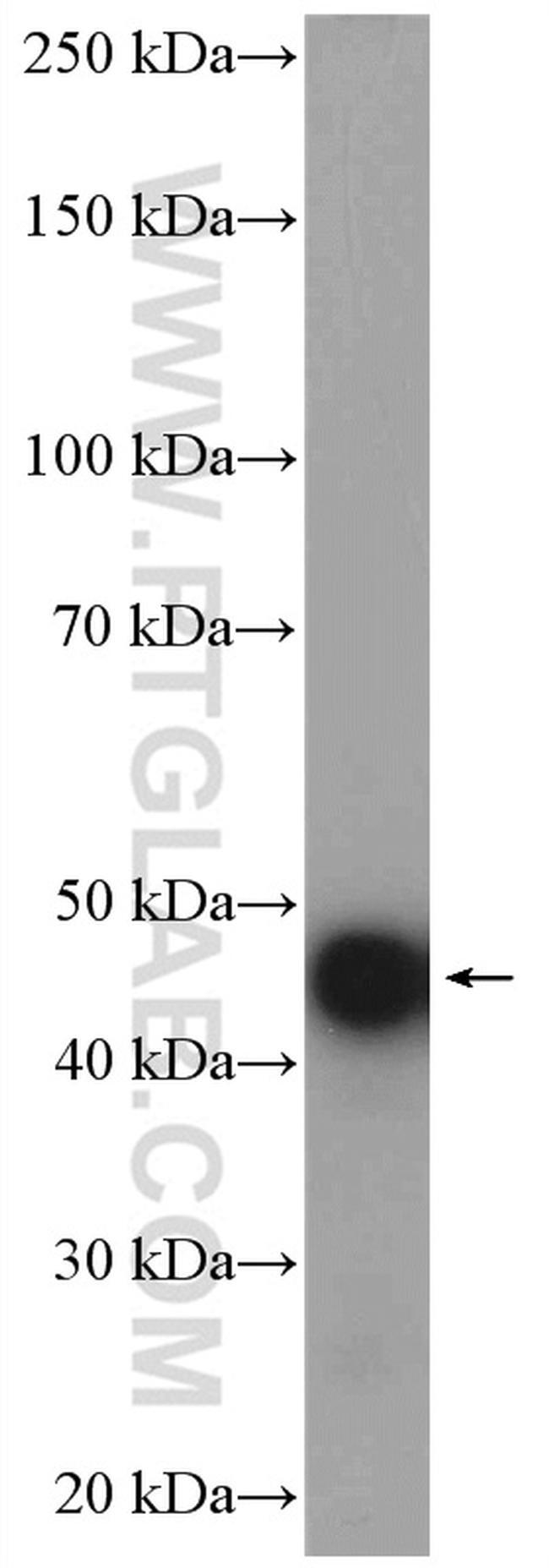
- Rat brain tissue were subjected to SDS PAGE followed by western blot with 12433-1-AP (HOMER1 antibody) at dilution of 1:5000 incubated at room temperature for 1 hour.
- Submitted by
- Invitrogen Antibodies (provider)
- Main image

- Experimental details
- NIH/3T3 cells were subjected to SDS PAGE followed by western blot with 12433-1-AP (HOMER1 antibody) at dilution of 1:1000 incubated at room temperature for 1.5 hours.

- Submitted by
- Invitrogen Antibodies (provider)
- Main image

- Experimental details
- C2C12 cell were subjected to SDS PAGE followed by western blot with 12433-1-AP (HOMER1 antibody) at dilution of 1:1000 incubated at room temperature for 1.5 hours.

Supportive validation
- Submitted by
- Invitrogen Antibodies (provider)
- Main image

- Experimental details
- Immunofluorescent analysis of ( -20°C Ethanol) fixed SH-SY5Y cells using 12433-1-AP (HOMER1 antibody) at dilution of 1:50 and Alexa Fluor 488-Conjugated AffiniPure Goat Anti-Rabbit IGG (H+L).

- Submitted by
- Invitrogen Antibodies (provider)
- Main image

- Experimental details
- Immunofluorescent analysis of ( -20°C Ethanol) fixed SH-SY5Y cells using 12433-1-AP (HOMER1 antibody) at dilution of 1:50 and Alexa Fluor 488-Conjugated AffiniPure Goat Anti-Rabbit IGG (H+L).

- Submitted by
- Invitrogen Antibodies (provider)
- Main image

- Experimental details
- Immunofluorescent analysis of (-20°C Ethanol) fixed MCF-7 cells using 12433-1-AP (HOMER1 antibody) at dilution of 1:50 and Alexa Fluor 488-conjugated AffiniPure Goat Anti-Rabbit IgG (H+L).

Supportive validation
- Submitted by
- Invitrogen Antibodies (provider)
- Main image
- Experimental details
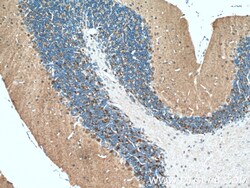
- Immunohistochemistry of paraffin-embedded mouse brain tissue slide using 12433-1-AP (HOMER1 antibody) at dilution of 1:200 (under 10x lens) heat mediated antigen retrieved with Tris-EDTA buffer (pH 9).

- Submitted by
- Invitrogen Antibodies (provider)
- Main image

- Experimental details
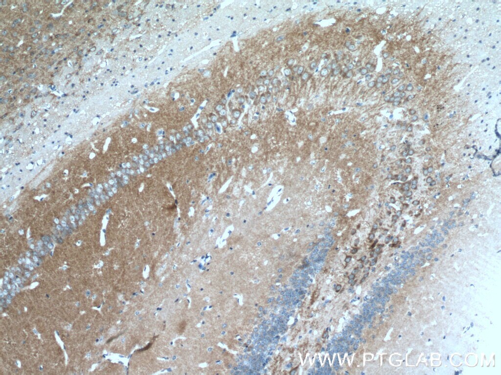
- Immunohistochemistry of paraffin-embedded mouse brain tissue slide using 12433-1-AP (HOMER1 antibody) at dilution of 1:200 (under 40x lens) heat mediated antigen retrieved with Tris-EDTA buffer (pH 9).

- Submitted by
- Invitrogen Antibodies (provider)
- Main image

- Experimental details
- Immunohistochemistry of paraffin-embedded mouse brain tissue slide using 12433-1-AP (HOMER1 antibody) at dilution of 1:200 (under 10x lens) heat mediated antigen retrieved with Tris-EDTA buffer (pH 9).
- Submitted by
- Invitrogen Antibodies (provider)
- Main image

- Experimental details
- Immunohistochemistry of paraffin-embedded mouse brain tissue slide using 12433-1-AP (HOMER1 antibody) at dilution of 1:200 (under 40x lens) heat mediated antigen retrieved with Tris-EDTA buffer (pH 9).

- Submitted by
- Invitrogen Antibodies (provider)
- Main image

- Experimental details
- Immunohistochemistry of paraffin-embedded human pancreas tissue slide using 12433-1-AP (HOMER1 Antibody) at dilution of 1:50 (under 10x lens).

- Submitted by
- Invitrogen Antibodies (provider)
- Main image

- Experimental details
- Immunohistochemistry of paraffin-embedded human pancreas tissue slide using 12433-1-AP (HOMER1 Antibody) at dilution of 1:50 (under 40x lens).

- Submitted by
- Invitrogen Antibodies (provider)
- Main image

- Experimental details
- Immunohistochemistry of paraffin-embedded human gliomas tissue slide using 12433-1-AP ( HOMER1 antibody) at dilution of 1:200 (under 10x lens) heat mediated antigen retrieved with Tris-EDTA buffer (pH 9).

- Submitted by
- Invitrogen Antibodies (provider)
- Main image

- Experimental details
- Immunohistochemistry of paraffin-embedded human gliomas tissue slide using 12433-1-AP ( HOMER1 antibody) at dilution of 1:200 (under 40x lens) heat mediated antigen retrieved with Tris-EDTA buffer (pH 9).

Supportive validation
- Submitted by
- Invitrogen Antibodies (provider)
- Main image
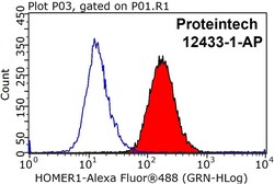
- Experimental details
- 1X10^6 HeLa cells were stained with 0.2ug HOMER1 antibody (12433-1-AP, red) and control antibody (blue). Fixed with 90% MeOH blocked with 3% BSA (30 min). Alexa Fluor 488-conjugated AffiniPure Goat Anti-Rabbit IGG (H+L) with dilution 1:1000.

Supportive validation
- Submitted by
- Invitrogen Antibodies (provider)
- Main image

- Experimental details
- IP result of anti-HOMER1 (IP:12433-1-AP, 4ug; Detection:12433-1-AP 1:1000) with fetal human brain tissue lysate 4000 ug.
