Explore
 Validate
 Learn
Learn Western blot
Antibody data
- Antibody Data
- Antigen structure
- References [1]
- Comments [0]
- Validations
- Western blot [1]
- Immunohistochemistry [1]
Submit
Validation data
Reference
Comment
Report error
- Product number
- MAB9166-100 - Provider product page

- Provider
- R&D Systems
- Product name
- Human/Mouse HSP47 Antibody
- Antibody type
- Monoclonal
- Description
- Protein A or G purified from hybridoma culture supernatant. Detects human and mouse HSP47 in direct ELISAs and Western blots.
- Reactivity
- Human, Mouse
- Host
- Mouse
- Conjugate
- Unconjugated
- Antigen sequence
P50454- Isotype
- IgG
- Antibody clone number
- 950806
- Vial size
- 100 ug
- Storage
- Use a manual defrost freezer and avoid repeated freeze-thaw cycles. 12 months from date of receipt, -20 to -70 °C as supplied. 1 month, 2 to 8 °C under sterile conditions after reconstitution. 6 months, -20 to -70 °C under sterile conditions after reconstitution.
Submitted references Crucial role of estrogen for the mammalian female in regulating semen coagulation and liquefaction in vivo.
Li S, Garcia M, Gewiss RL, Winuthayanon W
PLoS genetics 2017 Apr;13(4):e1006743
PLoS genetics 2017 Apr;13(4):e1006743
No comments: Submit comment
Supportive validation
- Submitted by
- R&D Systems (provider)
- Main image

- Experimental details
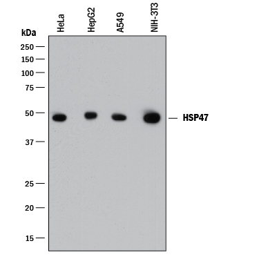
- Detection of Human and Mouse HSP47 by Western Blot. Western blot shows lysates of HeLa human cervical epithelial carcinoma cell line, HepG2 human hepatocellular carcinoma cell line, A549 human lung carcinoma cell line, and NIH-3T3 mouse embryonic fibroblast cell line. PVDF membrane was probed with 0.1 µg/mL of Mouse Anti-Human/Mouse HSP47 Monoclonal Antibody (Catalog # MAB9166) followed by HRP-conjugated Anti-Mouse IgG Secondary Antibody (Catalog # HAF018). A specific band was detected for HSP47 at approximately 47 kDa (as indicated). This experiment was conducted under reducing conditions and using Immunoblot Buffer Group 1.
Supportive validation
- Submitted by
- R&D Systems (provider)
- Main image
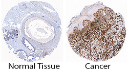
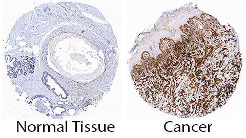
- Experimental details
- HSP47 in Human Breast Cancer Tissue. HSP47 was detected in immersion fixed paraffin-embedded sections of human normal breast (left) and breast cancer tissue (right) using Mouse Anti-Human/Mouse HSP47 Monoclonal Antibody (Catalog # MAB9166) at 15 µg/mL overnight at 4 °C. Tissue was stained using the Anti-Mouse HRP-DAB Cell & Tissue Staining Kit (brown; Catalog # CTS002) and counterstained with hematoxylin (blue). Specific staining was localized to cancer cell cytoplasm. View our protocol for Chromogenic IHC Staining of Paraffin-embedded Tissue Sections.