Explore
 Validate
 Learn
Learn Western blot
Antibody data
- Antibody Data
- Antigen structure
- References [0]
- Comments [0]
- Validations
- Western blot [4]
- Immunohistochemistry [1]
Submit
Validation data
Reference
Comment
Report error
- Product number
- PA1-26470 - Provider product page

- Provider
- Invitrogen Antibodies
- Product name
- Phospho-MYL9/MYL12 (Ser20) Polyclonal Antibody
- Antibody type
- Polyclonal
- Antigen
- Synthetic peptide
- Description
- Recommended positive controls: Cardiac myocytes.
- Concentration
- 1.19 mg/mL
No comments: Submit comment
Supportive validation
- Submitted by
- Invitrogen Antibodies (provider)
- Main image
- Experimental details
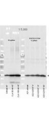
- Western blot analysis of Phospho-Myosin, Light chain pSer20 in 3 or 20 µL of a mouse cardiac myocyte lysate, either mock-treated or CLA-treated. Samples were loaded on a 4-20% Criterion gel for SDS-PAGE and probed with a Phospho-Myosin, Light chain pSer20 polyclonal antibody (Product # PA1-26470) at a dilution of 1:5000 and detected using an HRP conjugated Gt-anti-Rabbit IgG secondary antibody (color development using Amersham's substrate system).
- Submitted by
- Invitrogen Antibodies (provider)
- Main image
- Experimental details
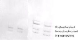
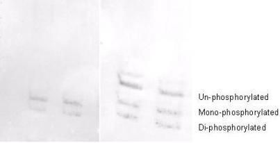
- Western blot analysis of Phospho-Myosin, Light chain pSer20 in NIH-3T3 cell lysates. Samples were probed with a Phospho-Myosin, Light chain pSer20 polyclonal antibody (Product # PA1-26470) at a dilution of 1:1000. A standard urea/glycerol gel without SDS was used to separate phospho forms of regulatory light chain according to mass to charge ratios. In Panel A, reactivity of the phosphospecific antibody is shown. In Panel B, reactivity of commercially available pan reactive antibody that detects both unphosphorylated and phosphorylated forms of regulatory light chain is shown. This phosphospecific antibody detects both monophosphorylated (pSer20 Mono-P-RLC) and diphosphorylated (pThr19-pSer20 Di-P-RLC) regulatory light chain.
- Submitted by
- Invitrogen Antibodies (provider)
- Main image

- Experimental details
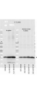
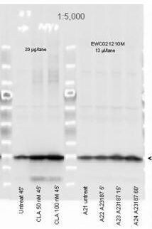
- Myosin Light chain (phospho Ser20) antibody Phospho-MYL9/MYL12 (Ser20) Polyclonal Antibody (Product # PA1-26470) was used at a 1:5,000 dilution to detect myosin light chain by Western blot. Either 13 or 20 µL of a mouse cardiac myocyte lysate was loaded on a 4-20% Criterion gel for SDS-PAGE. Samples were either mock-treated or CLA-treated, as indicated. After washing, a 1:5,000 dilution of HRP conjugated Gt-a-Rabbit IgG preceded color development using Amershams substrate system.
- Submitted by
- Invitrogen Antibodies (provider)
- Main image
- Experimental details
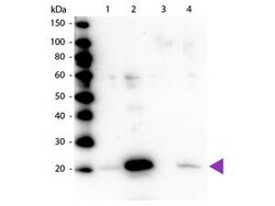
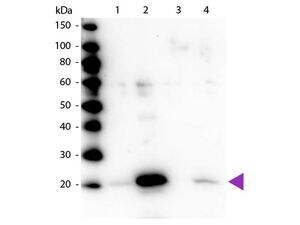
- Western blot analysis of Phospho-MYL9/MYL12 using Phospho-MYL9/MYL12 (Ser20) Polyclonal Antibody (Product # PA1-26470). Lane 1: Regulatory Light Chain Non-Phospho recombinant protein. Lane 2: Regulatory Light Chain Phospho recombinant protein. Lane 3: Smooth Muscle Non-Phospho recombinant protein. Lane 4: Smooth Muscle Phospho recombinant protein. Load: 50 ng per lane. Primary antibody at 1:1,000 overnight at 4°C. Secondary antibody: Peroxidase rabbit secondary antibody at 1:40,000 for 60 min at RT. Blocking for 30 min at RT. Predicted/Observed size: 20 kDa.
Supportive validation
- Submitted by
- Invitrogen Antibodies (provider)
- Main image

- Experimental details
- Immunohistochemistry analysis of Phospho-MYL9/MYL12 (Ser20) was performed in a variety of tissues including multi-human, multi-brain and multi-cancer slides using Phospho-MYL9/MYL12 (Ser20) Polyclonal Antibody (Product # PA1-26470) at 2.5 µg/mL. This image shows strong staining of both vascular and myometrial smooth muscle cells of the uterus. Tissue was formalin-fixed and paraffin embedded. The image shows localization of the antibody as the precipitated red signal, with a hematoxylin purple nuclear counterstain.