Antibody data
- Antibody Data
- Antigen structure
- References [0]
- Comments [0]
- Validations
- Western blot [1]
- Immunocytochemistry [2]
- Immunohistochemistry [3]
Submit
Validation data
Reference
Comment
Report error
- Product number
- TA307337 - Provider product page

- Provider
- OriGene
- Product name
- Anti-NRP1 Antibody [EPR3113]
- Antibody type
- Monoclonal
- Antigen
- A synthetic peptide corresponding to residues in the cytoplasmic region of human Neuropilin-1 was used as an immunogen.
- Description
- Rabbit monoclonal antibody against Neuropilin-1 (clone EPR3113 )
- Reactivity
- Human, Mouse, Rat, Simian
- Host
- Rabbit
- Isotype
- IgG
- Antibody clone number
- EPR3113
- Vial size
- 100 µl
No comments: Submit comment
Supportive validation
- Submitted by
- OriGene (provider)
- Main image

- Experimental details
- Western blot - Neuropilin 1 antibody [EPR3113]; All lanes : Anti-Neuropilin 1 antibody [EPR3113] at 1/1000 dilution.Lane 1 : Human placenta lysate.Lane 2 : HUVEC cell lysate.Lane 3 : HepG2 cell lysate.Lane 4 : mouse heart tissue lysate.Lane 5 : mouse kidney tissue lysate.Lane 6 : rat heart tissue lysate.Lane 7 : rat kidney tissue lysate.Lysates/proteins at 10 ug per lane.Secondary.HRP goat anti rabbit at 1/2000 dilution.Predicted band size : 103 kDa.Observed band size : 120 kDa .
- Validation comment
- WB
Supportive validation
- Submitted by
- OriGene (provider)
- Main image
- Experimental details
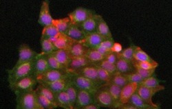
- ICC/IF image of TA307337 stained MCF7 cells. The cells were incubated with the antibody overnight at 4.. The secondary antibody (green) was DyLight 488 goat anti-rabbit IgG - H&L, pre-adsorbed used at 1:250. for 1h. Alexa Fluor 594 WGA was used to label plasma membranes (red) at 1:200 for 1h. DAPI was used to stain the cell nuclei (blue) at a concentration of 1.43uM.
- Validation comment
- IF
- Submitted by
- OriGene (provider)
- Main image
- Experimental details
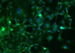
- ICC/IF - Anti-Neuropilin 1 antibody; TA307337 staining Neuropilin 1 in the COS1 fibroblast-like cell line derived from monkey kidney tissue by ICC/IF (ICC/IF). Cells were fixed with paraformaldehyde, permeabilized with Triton X-100 0.1% and blocked with 10% serum for 60 minutes at 24°C. Samples were incubated with primary antibody (1/200) for 16 hours at 4°C. An Alexa Fluor?488-conjugated Goat anti-rabbit monoclonal(1/500) was used as the secondary antibody.
- Validation comment
- IF
Supportive validation
- Submitted by
- OriGene (provider)
- Main image
- Experimental details
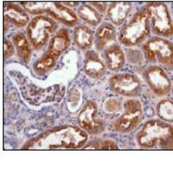
- Immunohistochemistry (Formalin/PFA-fixed paraffin-embedded sections) - Neuropilin 1 antibody [EPR3113]; Immunohistochemical analysis of paraffin-embedded human kidney tissue using TA307337 at 1/100 dilution.
- Validation comment
- IHC
- Submitted by
- OriGene (provider)
- Main image
- Experimental details
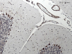
- IHC - Neuropilin 1 antibody; TA307337 staining Neuropilin 1 in Mouse brain tissue sections by IHC (IHC-P - formaldehyde-fixed, paraffin-embedded sections). Tissue was fixed with formaldehyde and blocked with 10% serum for 1 hour at RT; antigen retrieval was by heat mediation in citrate buffer (pH 6). Samples were incubated with primary antibody (1/100 in PBS + 2% blocking serum) for 16 hours at 4°C. A biotin-conjugated Goat anti-rabbit IgG polyclonal (1/250) was used as the secondary antibody.
- Validation comment
- IHC
- Submitted by
- OriGene (provider)
- Main image
- Experimental details
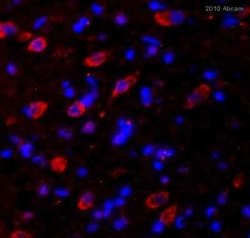
- Immunohistochemistry (Frozen sections) - Neuropilin 1 antibody [EPR3113]; TA307337 staining Neuropilin 1 in rat brain tissue sections by Immunohistochemistry (frozen sections). Tissue was fixed with paraformaldehyde and then blocked with 10% serum for 1 hour at 27?°C followed by incubation with the primary antibody, undiluted, for 14 hours at 4?°C. An undiluted Cy3?conjugated donkey anti-rabbit was used as the secondary antibody.
- Validation comment
- IHC
 Explore
 Validate
 Learn
Learn Western blot
 Flow cytometry