 Explore
 Validate
 Learn
Learn Western blot
 ELISA
ELISAAntibody data
- Antibody Data
- Antigen structure
- References [3]
- Comments [0]
- Validations
- Western blot [3]
- Immunohistochemistry [1]
- Flow cytometry [1]
Submit
Validation data
Reference
Comment
Report error
- Product number
- NB100-790 - Provider product page

- Provider
- Novus Biologicals
- Proper citation
- Novus Cat#NB100-790, RRID:AB_2235846
- Product name
- Goat Polyclonal NCF1 Antibody
- Antibody type
- Polyclonal
- Description
- Immunogen affinity purified.
- Reactivity
- Human, Mouse, Bacteria, Porcine
- Host
- Goat
- Isotype
- IgG
- Vial size
- 0.1 mg
- Concentration
- 0.5 mg/ml
- Storage
- Store at -20C. Avoid freeze-thaw cycles.
Submitted references Opportunistic Pathogen Porphyromonas gingivalis Modulates Danger Signal ATP-Mediated Antibacterial NOX2 Pathways in Primary Epithelial Cells.
Involvement of protein kinase D in Fc gamma-receptor activation of the NADPH oxidase in neutrophils.
Involvement of protein kinase D in Fc gamma-receptor activation of the NADPH oxidase in neutrophils.
Roberts JS, Atanasova KR, Lee J, Diamond G, Deguzman J, Hee Choi C, Yilmaz Ö
Frontiers in cellular and infection microbiology 2017;7:291
Frontiers in cellular and infection microbiology 2017;7:291
Involvement of protein kinase D in Fc gamma-receptor activation of the NADPH oxidase in neutrophils.
Davidson-Moncada JK, Lopez-Lluch G, Segal AW, Dekker LV
The Biochemical journal 2002 Apr 1;363(Pt 1):95-103
The Biochemical journal 2002 Apr 1;363(Pt 1):95-103
Involvement of protein kinase D in Fc gamma-receptor activation of the NADPH oxidase in neutrophils.
Davidson-Moncada JK, Lopez-Lluch G, Segal AW, Dekker LV
The Biochemical journal 2002 Apr 1;363(Pt 1):95-103
The Biochemical journal 2002 Apr 1;363(Pt 1):95-103
No comments: Submit comment
Supportive validation
- Submitted by
- Novus Biologicals (provider)
- Main image

- Experimental details
- Western Blot: NCF1 Antibody [NB100-790] - Staining of U937 lysate (35 ug protein in RIPA buffer). Primary incubation was 1 hour. Detected by chemiluminescence.

- Submitted by
- Novus Biologicals (provider)
- Main image

- Experimental details
- Western Blot: NCF1 Antibody [NB100-790] - Staining of Mouse Thymus (A) and Pig Spleen (B) lysate (35 ug protein in RIPA buffer). Antibody at 1 ug/mL. Primary incubation was 1 hour. Detected by chemiluminescence.

- Submitted by
- Novus Biologicals (provider)
- Main image

- Experimental details
- Western Blot: NCF1 Antibody [NB100-790] - Staining of U937 (A), Daudi (B), U251 (C) and negative control A431 (D) cell lysate (35 ug protein in RIPA buffer). Antibody at 0.2, 0.01, and 0.3 ug/mL respectively. Detected by chemiluminescence.

Supportive validation
- Submitted by
- Novus Biologicals (provider)
- Main image

- Experimental details
- Immunohistochemistry-Paraffin: NCF1 Antibody [NB100-790] - Staining of paraffin embedded Human Colon. Antibody at 5 ug/mL. Steamed antigen retrieval with citrate buffer pH 6, AP-staining.
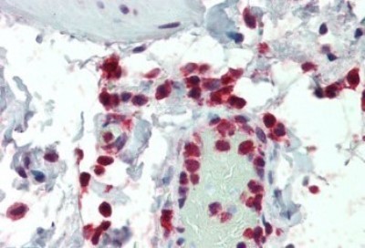
Supportive validation
- Submitted by
- Novus Biologicals (provider)
- Main image

- Experimental details
- Flow Cytometry: NCF1 Antibody [NB100-790] - Analysis of paraformaldehyde fixed HeLa cells (blue line), permeabilized with 0.5% Triton. Primary incubation 1hr (10 ug/mL) followed by Alexa Fluor 488 secondary antibody (1 ug/mL). IgG control: Unimmunized goat IgG (black line) followed by Alexa Fluor 488 secondary antibody.
