Explore
 Validate
 Learn
LearnPA5-20746
antibody from Invitrogen Antibodies
Targeting: KITLG
DFNA69, FPH2, Kitl, KL-1, MGF, SCF, SF, SLF
 Western blot
Antibody data
- Antibody Data
- Antigen structure
- References [2]
- Comments [0]
- Validations
- Western blot [5]
- Immunocytochemistry [1]
- Immunohistochemistry [2]
- Other assay [3]
Submit
Validation data
Reference
Comment
Report error
- Product number
- PA5-20746 - Provider product page

- Provider
- Invitrogen Antibodies
- Product name
- SCF Polyclonal Antibody
- Antibody type
- Polyclonal
- Antigen
- Synthetic peptide
- Description
- A suggested positive control is rat brain tissue lysate. PA5-20746 can be used with blocking peptide PEP-0860.
- Reactivity
- Human, Mouse, Rat
- Host
- Rabbit
- Isotype
- IgG
- Vial size
- 100 µg
- Concentration
- 1 mg/mL
- Storage
- Maintain refrigerated at 2-8°C for up to 3 months. For long term storage store at -20°C
Submitted references Schwann cells orchestrate peripheral nerve inflammation through the expression of CSF1, IL-34, and SCF in amyotrophic lateral sclerosis.
Cell Sheet Comprised of Mesenchymal Stromal Cells Overexpressing Stem Cell Factor Promotes Epicardium Activation and Heart Function Improvement in a Rat Model of Myocardium Infarction.
Trias E, Kovacs M, King PH, Si Y, Kwon Y, Varela V, Ibarburu S, Moura IC, Hermine O, Beckman JS, Barbeito L
Glia 2020 Jun;68(6):1165-1181
Glia 2020 Jun;68(6):1165-1181
Cell Sheet Comprised of Mesenchymal Stromal Cells Overexpressing Stem Cell Factor Promotes Epicardium Activation and Heart Function Improvement in a Rat Model of Myocardium Infarction.
Dergilev KV, Shevchenko EK, Tsokolaeva ZI, Beloglazova IB, Zubkova ES, Boldyreva MA, Menshikov MY, Ratner EI, Penkov D, Parfyonova YV
International journal of molecular sciences 2020 Dec 16;21(24)
International journal of molecular sciences 2020 Dec 16;21(24)
No comments: Submit comment
Supportive validation
- Submitted by
- Invitrogen Antibodies (provider)
- Main image
- Experimental details
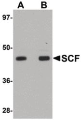
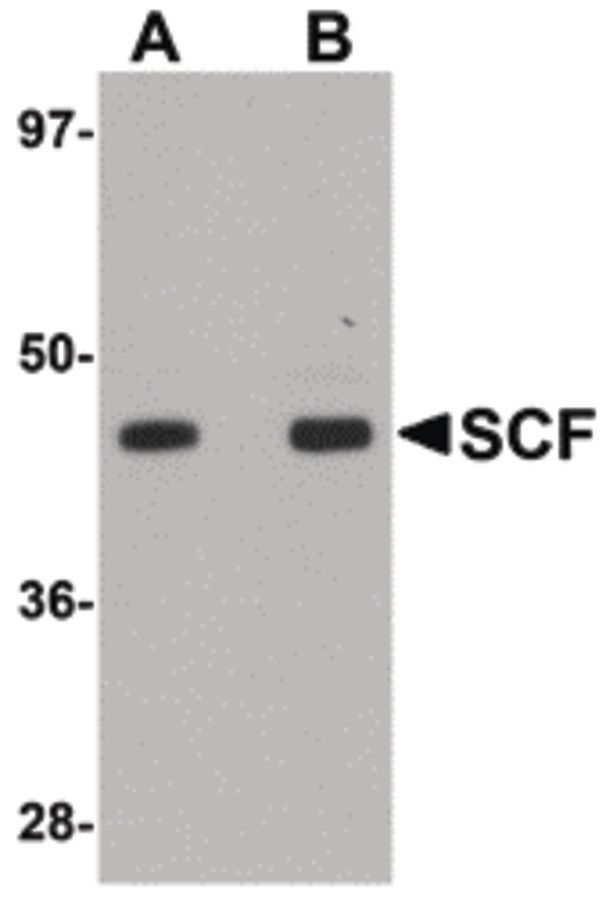
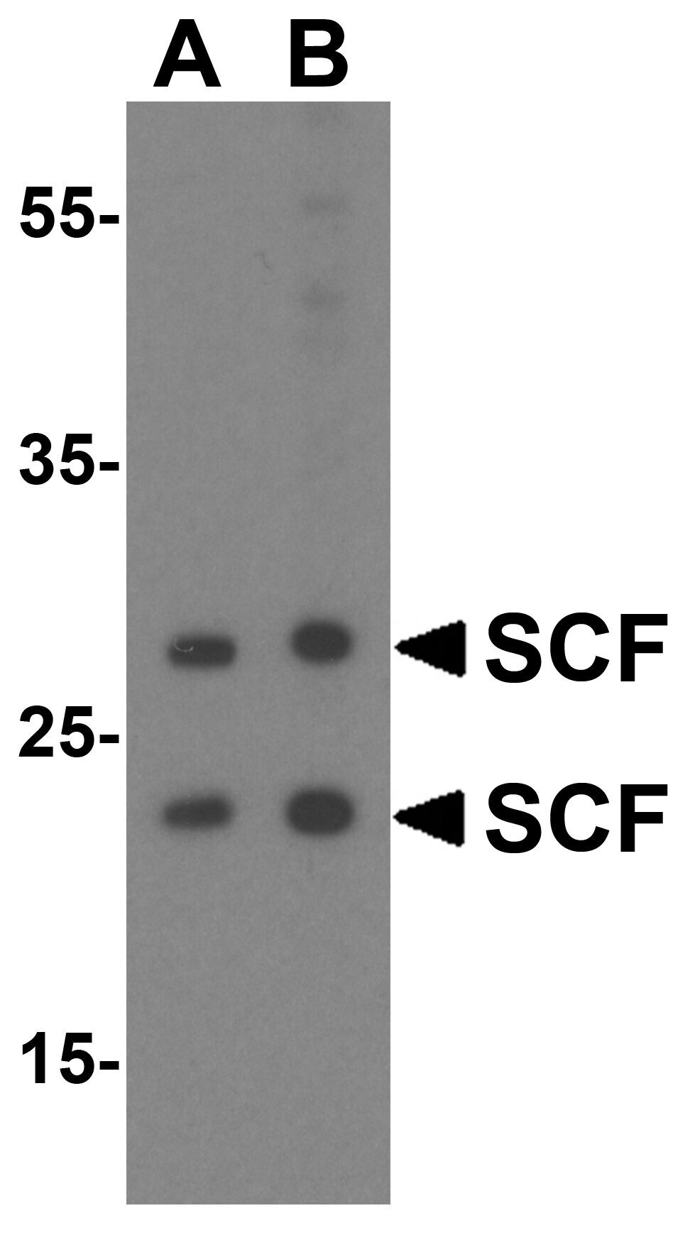
- Western blot analysis of rat brain tissue lysate using a SCF polyclonal antibody (Product # PA5-20746) at (A) 1 and (B) 2 µg/mL.
- Submitted by
- Invitrogen Antibodies (provider)
- Main image

- Experimental details
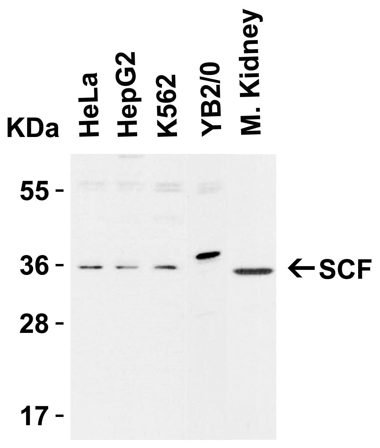
- Western Blot Validation in Cell Lines and Tissues of Human, Mouse and Rat. Loading: 15 µg of lysates per lane. Antibodies: SCF Polyclonal Antibody (Product # PA5-20746) (1 µg/mL), 1h incubation at RT in 0.05 NFDM/TBST. Secondary: Goat anti-rabbit IgG HRP conjugate at 1:10,000 dilution.

- Submitted by
- Invitrogen Antibodies (provider)
- Main image

- Experimental details
- Western Blot Validation in Cell Lines and Tissues of Human, Mouse and Rat. Loading: 15 µg of lysates per lane. Antibodies: SCF Polyclonal Antibody (Product # PA5-20746) (1 µg/mL), 1h incubation at RT in 0.05 NFDM/TBST. Secondary: Goat anti-rabbit IgG HRP conjugate at 1:10,000 dilution.
- Submitted by
- Invitrogen Antibodies (provider)
- Main image

- Experimental details
- Western Blot Validation in Human HeLa Cell Lines. Loading: 15 µg of lysates per lane. Antibodies: SCF Polyclonal Antibody (Product # PA5-20746) (2 µg/mL), 1h incubation at RT in 0.05 NFDM/TBST. Secondary: Goat anti-rabbit IgG HRP conjugate at 1:10,000 dilution.
- Submitted by
- Invitrogen Antibodies (provider)
- Main image

- Experimental details
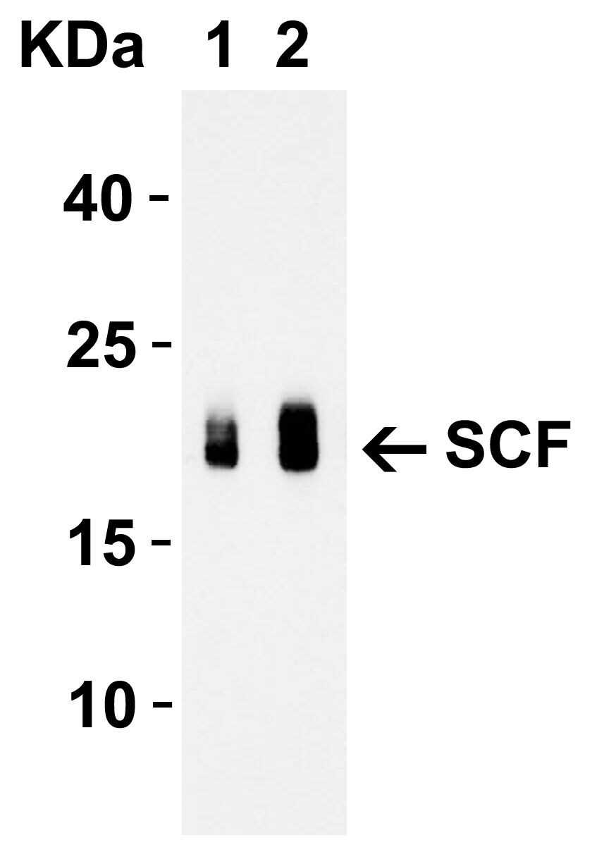
- Western Blot Validation with Recombinant Protein. Loading: 30 ng of human SCF recombinant protein per lane. Antibodies: SCF Polyclonal Antibody (Product # PA5-20746) (Lane 1: 1 µg/mL and Lane 2: 2 µg/mL), 1h incubation at RT in 0.05 NFDM/TBST. Secondary: Goat anti-rabbit IgG HRP conjugate at 1:10,000 dilution. Observed at around 20 kDa.
Supportive validation
- Submitted by
- Invitrogen Antibodies (provider)
- Main image
- Experimental details
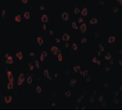
- Immunofluorescent analysis of human brain cells using a SCF polyclonal antibody (Product # PA5-20746) at a 20 µg/mL dilution.

Supportive validation
- Submitted by
- Invitrogen Antibodies (provider)
- Main image
- Experimental details
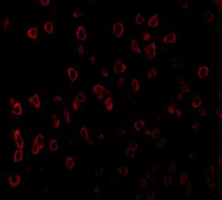
- Immunofluorescent analysis of 4% paraformaldehyde-fixed human brain tissue labeling SCF with SCF Polyclonal Antibody (Product # PA5-20746) at 20 µg/mL, followed by goat anti-rabbit IgG secondary antibody at 1:500 dilution (red).

- Submitted by
- Invitrogen Antibodies (provider)
- Main image
- Experimental details
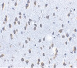
- Immunohistochemical analysis of paraffin-embedded mouse brain tissue using SCF Polyclonal Antibody (Product # PA5-20746) at 2.5 µg/mL. Tissue was fixed with formaldehyde and blocked with 0.1 serum for 1 h at RT; antigen retrieval was by heat mediation with a citrate buffer (pH6). Samples were incubated with primary antibody overnight at 4 ˚C. A goat anti-rabbit IgG H&L (HRP) at 1/250 was used as secondary. Counter stained with Hematoxylin.

Supportive validation
- Submitted by
- Invitrogen Antibodies (provider)
- Main image

- Experimental details
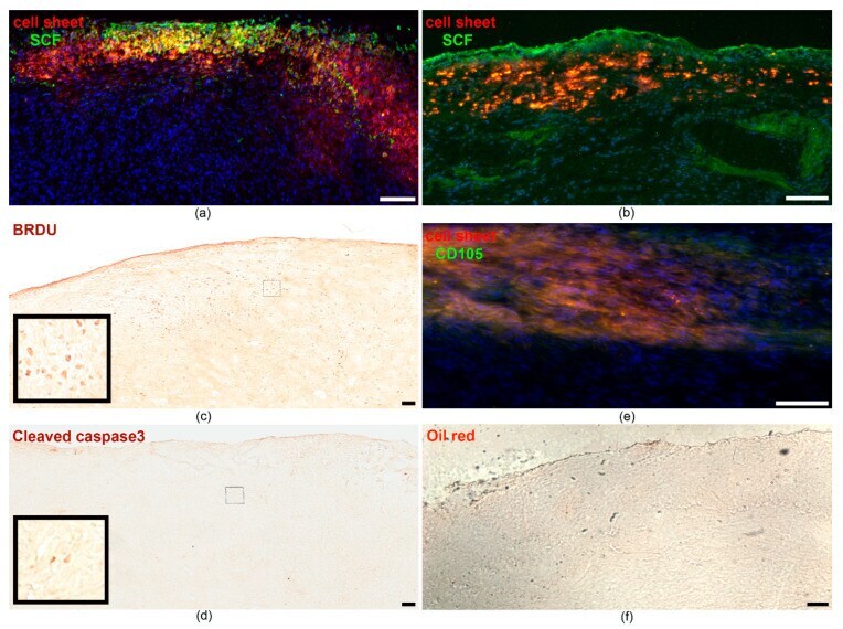
- Figure 3 SCF-MSC CS integration into myocardium tissue and transgene expression at day 5 ( a ) and 14 ( b ) after MI induction. Representative images of the infarction area that is covered by the cell sheet comprised of PKH26-labeled cells (red). Specific staining indicates SCF protein (green) distribution throughout the muscle tissue; ( c ) Bromodeoxyuridine (BRDU) immunohistostaining to identify proliferating cells in the graft and adjacent tissue (day 14). The small square bar indicate the area of the section where the magnified view (large square bar) was taken from; ( d ) Immunohistostaining for cleaved caspase 3 to identify apoptotic cells in the graft and adjacent tissue (day 14). The small square bar indicate the area of the section where the magnified view (large square bar) was taken from; ( e ) Immunofluorescent staining for CD105 (green) at day 14; ( f ) Oil-red staining to identify adipose cells (day 14). Nuclei are stained with DAPI (4', 6-diamidino-2-phenylindole). Scale bar-100 um.

- Submitted by
- Invitrogen Antibodies (provider)
- Main image

- Experimental details
- NULL

- Submitted by
- Invitrogen Antibodies (provider)
- Main image

- Experimental details
- Figure 3 SCF-MSC CS integration into myocardium tissue and transgene expression at day 5 ( a ) and 14 ( b ) after MI induction. Representative images of the infarction area that is covered by the cell sheet comprised of PKH26-labeled cells (red). Specific staining indicates SCF protein (green) distribution throughout the muscle tissue; ( c ) Bromodeoxyuridine (BRDU) immunohistostaining to identify proliferating cells in the graft and adjacent tissue (day 14). The small square bar indicate the area of the section where the magnified view (large square bar) was taken from; ( d ) Immunohistostaining for cleaved caspase 3 to identify apoptotic cells in the graft and adjacent tissue (day 14). The small square bar indicate the area of the section where the magnified view (large square bar) was taken from; ( e ) Immunofluorescent staining for CD105 (green) at day 14; ( f ) Oil-red staining to identify adipose cells (day 14). Nuclei are stained with DAPI (4', 6-diamidino-2-phenylindole). Scale bar-100 um.