Explore
 Validate
 Learn
LearnGTX22770
antibody from GeneTex
Targeting: AHR
bHLHe76
 Western blot
 ELISA Immunocytochemistry Immunoprecipitation Immunohistochemistry Chromatin Immunoprecipitation
Antibody data
- Antibody Data
- Antigen structure
- References [3]
- Comments [0]
- Validations
- Western blot [1]
- Immunocytochemistry [3]
- Immunohistochemistry [2]
Submit
Validation data
Reference
Comment
Report error
- Product number
- GTX22770 - Provider product page

- Provider
- GeneTex
- Proper citation
- GeneTex Cat#GTX22770, RRID:AB_384840
- Product name
- AHR antibody [RPT1]
- Antibody type
- Monoclonal
- Reactivity
- Human, Mouse, Rat, Porcine, Simian
- Host
- Mouse
Submitted references Functional expression of aryl hydrocarbon receptor on mast cells populating human endometriotic tissues.
Aryl hydrocarbon receptor-ligand axis mediates pulmonary fibroblast migration and differentiation through increased arachidonic acid metabolism.
Severe liver cirrhosis markedly reduces AhR-mediated induction of cytochrome P450 in rats by decreasing the transcription of target genes.
Mariuzzi L, Domenis R, Orsaria M, Marzinotto S, Londero AP, Bulfoni M, Candotti V, Zanello A, Ballico M, Mimmi MC, Calcagno A, Marchesoni D, Di Loreto C, Beltrami AP, Cesselli D, Gri G
Laboratory investigation; a journal of technical methods and pathology 2016 Sep;96(9):959-971
Laboratory investigation; a journal of technical methods and pathology 2016 Sep;96(9):959-971
Aryl hydrocarbon receptor-ligand axis mediates pulmonary fibroblast migration and differentiation through increased arachidonic acid metabolism.
Su HH, Lin HT, Suen JL, Sheu CC, Yokoyama KK, Huang SK, Cheng CM
Toxicology 2016 Aug 31;370:116-126
Toxicology 2016 Aug 31;370:116-126
Severe liver cirrhosis markedly reduces AhR-mediated induction of cytochrome P450 in rats by decreasing the transcription of target genes.
Floreani M, De Martin S, Gabbia D, Barbierato M, Nassi A, Mescoli C, Orlando R, Bova S, Angeli P, Gola E, Sticca A, Palatini P
PloS one 2013;8(4):e61983
PloS one 2013;8(4):e61983
No comments: Submit comment
Supportive validation
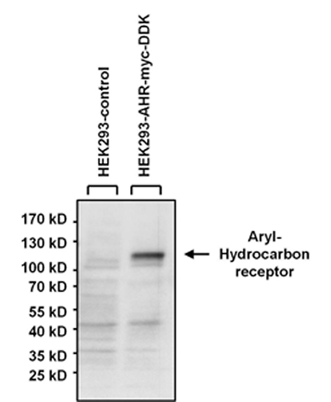
- Submitted by
- GeneTex (provider)
- Main image

- Experimental details
- Western blot analysis of Aryl Hydrocarbon Receptor was performed by loading 40ug of HEK293 lysate overexpressing Aryl Hydrocarbon Receptor (right lane) or empty vector control (left lane) and 10ul of prestained protein ladder onto a 4-20% Tris-HCl polyacrylamide gel. Proteins were transferred to a PVDF membrane and blocked with 5% BSA/TBST for at least 1 hour. The membrane was probed with an Aryl Hydrocarbon Receptor monoclonal antibody (GTX22770) at a dilution of 1:1000 overnight at 4?C on a rocking platform, washed in TBS-0.1%Tween-20, and probed with a goat anti-mouse IgG-HRP secondary antibody at a dilution of 1:20,000 for 1 hour. Chemiluminescent detection was performed
Supportive validation
- Submitted by
- GeneTex (provider)
- Main image
- Experimental details
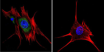
- Immunofluorescent analysis of Aryl Hydrocarbon Receptor in HeLa cells. Cells were grown on chamber slides and fixed with formaldehyde prior to staining. Cells were probed without (control) or with a Aryl Hydrocarbon Receptor monoclonal antibody (GTX22770) at a dilution of 1:200 overnight at 4 C, washed with PBS and incubated with a DyLight-488 conjugated secondary antibody. Aryl Hydrocarbon Receptor staining (green), F-Actin staining with Phalloidin (red) and nuclei with DAPI (blue) is shown.

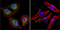
- Submitted by
- GeneTex (provider)
- Main image
- Experimental details
- Immunofluorescent analysis of Aryl Hydrocarbon Receptor in MCF-7 cells. Cells were grown on chamber slides and fixed with formaldehyde prior to staining. Cells were probed without (control) or with a Aryl Hydrocarbon Receptor monoclonal antibody (GTX22770) at a dilution of 1:100 overnight at 4 C, washed with PBS and incubated with a DyLight-488 conjugated secondary antibody. Aryl Hydrocarbon Receptor staining (green), F-Actin staining with Phalloidin (red) and nuclei with DAPI (blue) is shown.

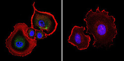
- Submitted by
- GeneTex (provider)
- Main image

- Experimental details
- Immunofluorescent analysis of Aryl Hydrocarbon Receptor in NIH-3T3 cells. Cells were grown on chamber slides and fixed with formaldehyde prior to staining. Cells were probed without (control) or with a Aryl Hydrocarbon Receptor monoclonal antibody (GTX22770) at a dilution of 1:200 overnight at 4 C, washed with PBS and incubated with a DyLight-488 conjugated secondary antibody. Aryl Hydrocarbon Receptor staining (green), F-Actin staining with Phalloidin (red) and nuclei with DAPI (blue) is shown.
Supportive validation
- Submitted by
- GeneTex (provider)
- Main image
- Experimental details
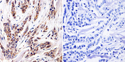
- Immunohistochemistry was performed on cancer biopsies of de-paraffinized human breast carcinoma tissue. To expose target proteins, heat induced antigen retrieval was performed using 10mM sodium citrate (pH 6.0) buffer, microwaved for 8-15 minutes. Following antigen retrieval tissues were blocked in 3% BSA-PBS for 30 minutes at room temperature and probed with a Aryl Hydrocarbon Receptor monoclonal antibody (GTX22770) at a dilution of 1:100 or without primary antibody (negative control) overnight at 4¢XC in a humidified chamber. Tissues were washed with PBST and endogenous peroxidase activity was quenched with a peroxidase suppressor. Detection was performed using a biotin-conjugated secondary antibody and SA-HRP, followed by colorimetric detection using DAB. Tissues were counterstained with hematoxylin and prepped for mounting.

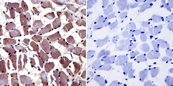
- Submitted by
- GeneTex (provider)
- Main image

- Experimental details
- Immunohistochemistry was performed on normal biopsies of de-paraffinized human skeletal muscle tissue. To expose target proteins, heat induced antigen retrieval was performed using 10mM sodium citrate (pH 6.0) buffer, microwaved for 8-15 minutes. Following antigen retrieval tissues were blocked in 3% BSA-PBS for 30 minutes at room temperature and probed with a Aryl Hydrocarbon Receptor monoclonal antibody (GTX22770) at a dilution of 1:20 or without primary antibody (negative control) overnight at 4¢XC in a humidified chamber. Tissues were washed with PBST and endogenous peroxidase activity was quenched with a peroxidase suppressor. Detection was performed using a biotin-conjugated secondary antibody and SA-HRP, followed by colorimetric detection using DAB. Tissues were counterstained with hematoxylin and prepped for mounting.