 Explore
 Validate
 Learn
LearnMA5-16318
antibody from Invitrogen Antibodies
Targeting: CA9
CAIX, MN
 Western blot Immunocytochemistry
 Immunoprecipitation Immunohistochemistry Flow cytometry Other assay
Antibody data
- Antibody Data
- Antigen structure
- References [3]
- Comments [0]
- Validations
- Western blot [2]
- Immunocytochemistry [1]
- Immunohistochemistry [4]
- Flow cytometry [1]
- Other assay [2]
Submit
Validation data
Reference
Comment
Report error
- Product number
- MA5-16318 - Provider product page

- Provider
- Invitrogen Antibodies
- Product name
- Carbonic Anhydrase IX Monoclonal Antibody (GT12)
- Antibody type
- Monoclonal
- Antigen
- Other
- Reactivity
- Human
- Host
- Mouse
- Isotype
- IgG
- Antibody clone number
- GT12
- Vial size
- 100 µL
- Concentration
- 0.1 mg/mL
- Storage
- Store at 4°C short term. For long term storage, store at -20°C, avoiding freeze/thaw cycles.
Submitted references Hypoxia-induced autophagy drives colorectal cancer initiation and progression by activating the PRKC/PKC-EZR (ezrin) pathway.
Linker stability influences the anti-tumor activity of acetazolamide-drug conjugates for the therapy of renal cell carcinoma.
Mineralization of bone-related SaOS-2 cells under physiological hypoxic conditions.
Qureshi-Baig K, Kuhn D, Viry E, Pozdeev VI, Schmitz M, Rodriguez F, Ullmann P, Koncina E, Nurmik M, Frasquilho S, Nazarov PV, Zuegel N, Boulmont M, Karapetyan Y, Antunes L, Val D, Mittelbronn M, Janji B, Haan S, Letellier E
Autophagy 2020 Aug;16(8):1436-1452
Autophagy 2020 Aug;16(8):1436-1452
Linker stability influences the anti-tumor activity of acetazolamide-drug conjugates for the therapy of renal cell carcinoma.
Cazzamalli S, Corso AD, Neri D
Journal of controlled release : official journal of the Controlled Release Society 2017 Jan 28;246:39-45
Journal of controlled release : official journal of the Controlled Release Society 2017 Jan 28;246:39-45
Mineralization of bone-related SaOS-2 cells under physiological hypoxic conditions.
Müller WE, Schröder HC, Tolba E, Diehl-Seifert B, Wang X
The FEBS journal 2016 Jan;283(1):74-87
The FEBS journal 2016 Jan;283(1):74-87
No comments: Submit comment
Supportive validation
- Submitted by
- Invitrogen Antibodies (provider)
- Main image

- Experimental details
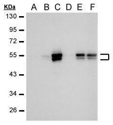
- Western Blot analysis of Carbonic Anhydrase IX was performed by separating 30 µg of various whole cell extracts by SDS PAGE. Proteins were transferred to a membrane and probed with a Carbonic Anhydrase IX Monoclonal Antibody (GT12) (Product # MA5-16318) at a dilution of 1:1000. The HRP-conjugated anti-mouse IgG antibody was used to detect the primary antibody. A: 24 hr Untreated, B: 24 hr treatment with 100µM CoCl, C:24 hr treatment with200µM CoCl, D: 48 hr Untreated, E: 48 hr treatment with 100 µM CoCl, F: 48 hr treatment with 200 µM CoCl.
- Submitted by
- Invitrogen Antibodies (provider)
- Main image

- Experimental details
- Western blot was performed using Anti-Carbonic Anhydrase IX Monoclonal Antibody (Product # MA5-16318) and ~ 50-55 kDa bands corresponding to CA9 was observed across cell lines tested and increased upon Deferoxamine and Cobalt Chloride treatment in HeLa along with an uncharacterized band (*) at ~32 kDa. Whole cell extracts (30 µg lysate) of U-87 MG (Lane 1), HeLa (Lane 2), HeLa treated with Deferoxamine (100uM for 24 Hours) (Lane 3) and HeLa treated with Cobalt Chloride (200uM for 24 Hours) (Lane 4) were electrophoresed using NuPAGE® 4-12 % Bis-Tris gel (Product # NP0322BOX). Resolved proteins were then transferred onto a nitrocellulose membrane (Product # IB23001) by iBlot® 2 Dry Blotting System (Product # IB21001). The blot was probed with the primary antibody (1:500 dilution) and detected by chemiluminescence with Goat anti-Mouse IgG (H+L), Superclonal™ Recombinant Secondary Antibody, HRP (Product # A28177, 1:4000 dilution) using the iBright FL 1000 (Product # A32752). Chemiluminescent detection was performed using Novex® ECL Chemiluminescent Substrate Reagent Kit (Product # WP20005).

Supportive validation
- Submitted by
- Invitrogen Antibodies (provider)
- Main image
- Experimental details
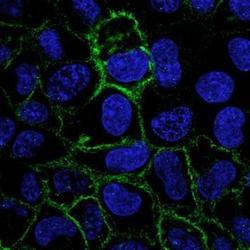
- Confocal immunofluorescence analysis (Olympus FV10i) of methanol-fixed A431 cells treated with 200 µM CoCl2 for 48hr using anti-CAIX antibody [GT12] (Product # MA5-16318) at a dilution of 1:1,000.
Supportive validation
- Submitted by
- Invitrogen Antibodies (provider)
- Main image

- Experimental details
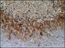
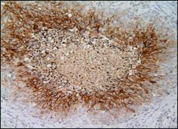
- Immunohistochemistry (Paraffin) analysis of Carbonic Anhydrase IX was performed in paraffin-embedded renal cell carcinoma (clear cell type) tissue using Carbonic Anhydrase IX Monoclonal Antibody (GT12) (Product # MA5-16318) at a dilution of 1:1000.

- Submitted by
- Invitrogen Antibodies (provider)
- Main image

- Experimental details
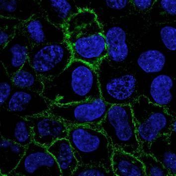
- Carbonic Anhydrase IX Monoclonal Antibody (GT12) detects Carbonic Anhydrase IX protein at cell membrane by immunohistochemical analysis. Sample: Paraffin-embedded human cervical carcinoma. Carbonic Anhydrase IX stained by Carbonic Anhydrase IX Monoclonal Antibody (GT12) (Product # MA5-16318) diluted at 1:500. Antigen Retrieval: Citrate buffer, pH 6.0, 15 min.

- Submitted by
- Invitrogen Antibodies (provider)
- Main image
- Experimental details
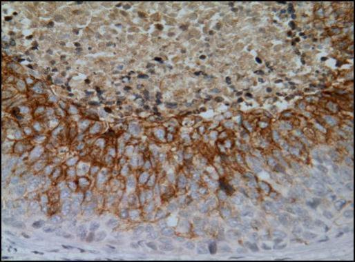
- Immunohistochemical analysis of paraffin-embedded cervical CA tissue sections using anti-CAIX antibody [GT12] (Product # MA5-16318) at a dilution of 1:1,000. The hypoxic regions of the tumor show positive CAIX staining.
- Submitted by
- Invitrogen Antibodies (provider)
- Main image
- Experimental details
- Immunohistochemical analysis of paraffin-embedded cervical CA tissue sections using anti-CAIX antibody [GT12] (Product # MA5-16318) at a dilution of 1:1,000. The hypoxic regions of the tumor show positive CAIX staining.

Supportive validation
- Submitted by
- Invitrogen Antibodies (provider)
- Main image
- Experimental details
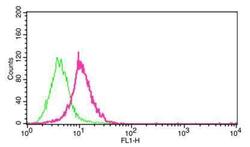
- Flow cytometry on HeLa cells (1x10^6) stained with Carbonic Anhydrase IX Monoclonal Antibody (GT12) (Product # MA5-16318) at a 1:1,000 dilution. HeLa cells were untreated (green) or treated with 200 µM CoCl2 (pink) for 48 hr.
Supportive validation
- Submitted by
- Invitrogen Antibodies (provider)
- Main image
- Experimental details
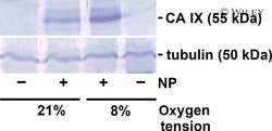
- NULL

- Submitted by
- Invitrogen Antibodies (provider)
- Main image

- Experimental details
- Figure 7. The relevance of the hypoxia/autophagy/EZR pathway in human TICs. ( A ) Immunofluorescence staining and colocalization in human tumor tissues (from six patients) for CA9 (a downstream target of HIF1A), BNIP3L, MAP1LC3A, p-EZR, and POU5F1 (refer to Fig. S7 for patient characteristics including TNM staging and HE stainings). The specificity of all used antibodies was carefully validated (please see Material and Methods and Supplementary data). Scale bar: 100 um. ( B ) Staining correlation in human CRC tissues. Measures were standardized (z-score) for each patient. Dot colors indicate different patients. A repeated measure correlation test was performed in order to account for the within-individual association of paired measures (using the rmcorr package in R; see Material and Methods). The rmcorr r coefficient and the Holm adjusted p-values are reported on each plot.
