Explore
 Validate
 Learn
Learn Western blot
Antibody data
- Antibody Data
- Antigen structure
- References [0]
- Comments [0]
- Validations
- Western blot [1]
- Immunocytochemistry [1]
- Other assay [4]
Submit
Validation data
Reference
Comment
Report error
- Product number
- H00003055-D01P - Provider product page

- Provider
- Invitrogen Antibodies
- Product name
- HCK Polyclonal Antibody, MaxPab™
- Antibody type
- Polyclonal
- Antigen
- Other
- Reactivity
- Human
- Host
- Rabbit
- Isotype
- IgG
- Vial size
- 100 µg
- Concentration
- See Label
- Storage
- -20° C, Avoid Freeze/Thaw Cycles
No comments: Submit comment
Supportive validation
- Submitted by
- Invitrogen Antibodies (provider)
- Main image
- Experimental details
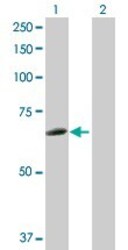
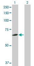
- Western Blot analysis of HCK expression in transfected 293T cell line (H00003055-T02) by HCK MaxPab polyclonal antibody.Lane 1: HCK transfected lysate(57.30 KDa).Lane 2: Non-transfected lysate.
Supportive validation
- Submitted by
- Invitrogen Antibodies (provider)
- Main image
- Experimental details
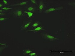
- Immunofluorescence of purified MaxPab antibody to HCK on HeLa cell. Antibody concentration is 10 µg/mL.

Supportive validation
- Submitted by
- Invitrogen Antibodies (provider)
- Main image
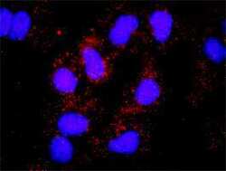
- Experimental details
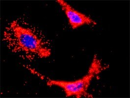
- Proximity Ligation Analysis of protein-protein interactions between HCK and SOS1. Huh7 cells were stained with anti-HCK rabbit purified polyclonal 1:1200 and anti-SOS1 mouse monoclonal antibody 1:50. Each red dot represents the detection of protein-protein interaction complex, and nuclei were counterstained with DAPI (blue).

- Submitted by
- Invitrogen Antibodies (provider)
- Main image

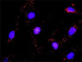
- Experimental details
- Proximity Ligation Analysis of protein-protein interactions between HCK and CRKL. Mahlavu cells were stained with anti-HCK rabbit purified polyclonal 1:1200 and anti-CRKL mouse monoclonal antibody 1:50. Each red dot represents the detection of protein-protein interaction complex, and nuclei were counterstained with DAPI (blue).
- Submitted by
- Invitrogen Antibodies (provider)
- Main image

- Experimental details
- Proximity Ligation Analysis of protein-protein interactions between HCK and SOS1. Huh7 cells were stained with anti-HCK rabbit purified polyclonal 1:1200 and anti-SOS1 mouse monoclonal antibody 1:50. Each red dot represents the detection of protein-protein interaction complex, and nuclei were counterstained with DAPI (blue).
- Submitted by
- Invitrogen Antibodies (provider)
- Main image

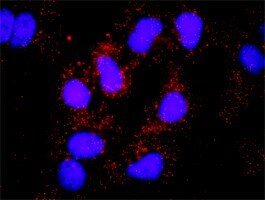
- Experimental details
- Proximity Ligation Analysis of protein-protein interactions between HCK and PIK3CB. HeLa cells were stained with anti-HCK rabbit purified polyclonal 1:1200 and anti-PIK3CB mouse monoclonal antibody 1:50. Each red dot represents the detection of protein-protein interaction complex, and nuclei were counterstained with DAPI (blue).