Explore
 Validate
 Learn
Learn Western blot
Antibody data
- Antibody Data
- Antigen structure
- References [0]
- Comments [0]
- Validations
- Western blot [2]
- Immunocytochemistry [1]
- Immunohistochemistry [1]
- Flow cytometry [1]
Submit
Validation data
Reference
Comment
Report error
- Product number
- TA324961 - Provider product page

- Provider
- OriGene
- Product name
- Rabbit polyclonal FGFR4 Antibody (N-term)
- Antibody type
- Polyclonal
- Description
- Rabbit polyclonal FGFR4 Antibody (N-term)
- Host
- Rabbit
- Conjugate
- Unconjugated
- Epitope
- FGFR4
- Isotype
- IgG
- Antibody clone number
- NULL
- Vial size
- 400 µl
- Concentration
- 0.5 mg/ml
No comments: Submit comment
Supportive validation
- Submitted by
- OriGene (provider)
- Main image
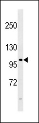
- Experimental details
- FGFR4 Antibody (E39) (Cat. #TA324961) western blot analysis in 293 cell line lysates (35ug/lane).This demonstrates the FGFR4 antibody detected the FGFR4 protein (arrow).
- Validation comment
- WB
- Submitted by
- OriGene (provider)
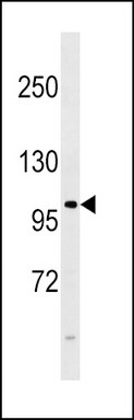
- Main image
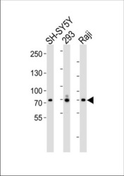
- Experimental details
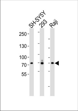
- Western blot analysis of lysates from SH-SY5Y 293, Raji cell line (from left to right), using FGFR4 Antibody (N-term) (Cat. #TA324961). TA324961 was diluted at 1:1000 at each lane. A goat anti-rabbit IgG H&L(HRP) at 1:5000 dilution was used as the secondary antibody. Lysates at 35ug per lane.
- Validation comment
- WB
Supportive validation
- Submitted by
- OriGene (provider)
- Main image
- Experimental details
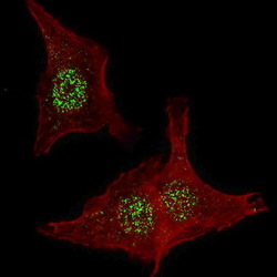
- IF image of HeLa cells stained with FGFR4 (N-term) antibody. HeLa cells were incubated with TA324961 FGFR4 (N-term) primary antibody (1:200, 2 h at RT). For secondary antibody, Alexa Fluor? 488 conjugated donkey anti-rabbit antibody (green) was used (1:1000, 1h). Nuclei were counterstained with Hoechst 33342 (blue) . Note the highly specific localization of the FGFR4 mainly to the nucleus, supported by Human Protein Atlas Data (http://www.proteinatlas.org/ENSG00000160867).
- Validation comment
- IF
Supportive validation
- Submitted by
- OriGene (provider)
- Main image

- Experimental details
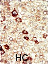
- Formalin-fixed and paraffin-embedded human cancer tissue reacted with the primary antibody, which was peroxidase-conjugated to the secondary antibody, followed by AEC staining. This data demonstrates the use of this antibody for immunohistochemistry; clinical relevance has not been evaluated. BC = breast carcinoma; HC = hepatocarcinoma.
- Validation comment
- IHC
Supportive validation
- Submitted by
- OriGene (provider)
- Main image
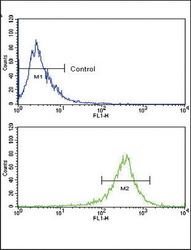
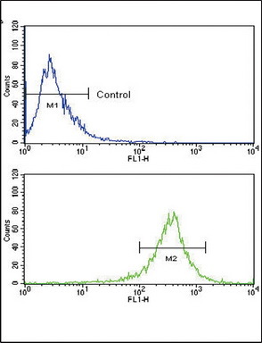
- Experimental details
- Flow cytometric analysis of WiDr cells using FGFR4 Antibody (N-term) (bottom histogram) compared to a negative control cell (top histogram). FITC-conjugated goat-anti-rabbit secondary antibodies were used for the analysis.
- Validation comment
- FC