 Explore
 Validate
 Learn
Learn Western blot
 Immunocytochemistry
Antibody data
- Antibody Data
- Antigen structure
- References [2]
- Comments [0]
- Validations
- Western blot [7]
- Immunocytochemistry [1]
- Immunoprecipitation [1]
- Immunohistochemistry [4]
Submit
Validation data
Reference
Comment
Report error
- Product number
- GTX102580 - Provider product page

- Provider
- GeneTex
- Proper citation
- GeneTex Cat#GTX102580, RRID:AB_2037126
- Product name
- Grp78 antibody [N2C1], Internal
- Antibody type
- Polyclonal
- Reactivity
- Human, Mouse, Rat
- Host
- Rabbit
Submitted references The role of sodium hydrosulfide in attenuating the aging process via PI3K/AKT and CaMKKβ/AMPK pathways.
Periodontal disease level-butyric acid amounts locally administered in the rat gingival mucosa induce ER stress in the systemic blood.
Chen X, Zhao X, Cai H, Sun H, Hu Y, Huang X, Kong W, Kong W
Redox biology 2017 Aug;12:987-1003
Redox biology 2017 Aug;12:987-1003
Periodontal disease level-butyric acid amounts locally administered in the rat gingival mucosa induce ER stress in the systemic blood.
Cueno ME, Saito Y, Ochiai K
Microbial pathogenesis 2016 May;94:70-5
Microbial pathogenesis 2016 May;94:70-5
No comments: Submit comment
Enhanced validation
Supportive validation
- Submitted by
- GeneTex (provider)
- Enhanced method
- Genetic validation
- Main image

- Experimental details
- Non-transfected (¡V) and transfected (+) HepG2 whole cell extracts (30 ?g) were separated by 7.5% SDS-PAGE, and the membrane was blotted with Grp78 antibody [N2C1], Internal (GTX102580) diluted at 1:10000. The HRP-conjugated anti-rabbit IgG antibody (GTX213110-01) was used to detect the primary antibody.

Supportive validation
- Submitted by
- GeneTex (provider)
- Main image

- Experimental details
- Grp78 antibody [N2C1], Internal detects Grp78 protein by western blot analysis.A. 30 ?g 293T whole cell lysate/extract B. 30 ?g A431 whole cell lysate/extract C. 30 ?g HeLa whole cell lysate/extract D. 30 ?g HepG2 whole cell lysate/extract7.5 % SDS-PAGEGrp78 antibody [N2C1], Internal (GTX102580) dilution: 1:5000
- Validation comment
- WB

- Submitted by
- GeneTex (provider)
- Main image

- Experimental details
- Grp78 antibody detects Grp78 protein by western blot analysis. Various whole cell extracts (30 ?g) were separated by 7.5% SDS-PAGE, and the membrane was blotted with Grp78 antibody (GTX102580) diluted at a dilution of 1:5000.
- Validation comment
- WB

- Submitted by
- GeneTex (provider)
- Main image

- Experimental details
- Grp78 antibody detects Grp78 protein by western blot analysis. Various whole cell extracts (30 ?g) were separated by 7.5% SDS-PAGE, and the membrane was blotted with Grp78 antibody (GTX102580) diluted at a dilution of 1:5000. The HRP-conjugated anti-rabbit IgG antibody (GTX213110-01) was used to detect the primary antibody.

- Submitted by
- GeneTex (provider)
- Main image

- Experimental details
- Various whole cell extracts (30 ?g) were separated by 7.5% SDS-PAGE, and the membrane was blotted with Grp78 antibody [N2C1], Internal (GTX102580) diluted at 1:5000. The HRP-conjugated anti-rabbit IgG antibody (GTX213110-01) was used to detect the primary antibody.

- Submitted by
- GeneTex (provider)
- Main image

- Experimental details
- Non-transfected (¡V) and transfected (+) HepG2 whole cell extracts (30 ?g) were separated by 7.5% SDS-PAGE, and the membrane was blotted with Grp78 antibody [N2C1], Internal (GTX102580) diluted at 1:10000. The HRP-conjugated anti-rabbit IgG antibody (GTX213110-01) was used to detect the primary antibody.

Supportive validation
- Submitted by
- GeneTex (provider)
- Main image

- Experimental details
- Grp78 antibody [N2C1], Internal detects Grp78 protein at endoplasmic reticulum by immunofluorescent analysis.Sample: HeLa cells were fixed in ice-cold MeOH for 5 min.Green: Grp78 protein stained by Grp78 antibody [N2C1], Internal (GTX102580) diluted at 1:500.Blue: Hoechst 33342 staining.

Supportive validation
- Submitted by
- GeneTex (provider)
- Main image
- Experimental details
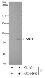
- Immunoprecipitation of Grp78 protein from HepG2 whole cell extracts using 5 £gg of Grp78 antibody [N2C1], Internal (GTX102580).Western blot analysis was performed using Grp78 antibody [N2C1], Internal (GTX102580).EasyBlot anti-Rabbit IgG (GTX221666-01) was used as a secondary reagent.

Supportive validation
- Submitted by
- GeneTex (provider)
- Main image

- Experimental details
- Grp78 antibody [N2C1], Internal detects Grp78 protein at cytosol on mouse intestine by immunohistochemical analysis. Sample: Paraffin-embedded mouse intestine. Grp78 antibody [N2C1], Internal (GTX102580) dilution: 1:500.

- Submitted by
- GeneTex (provider)
- Main image

- Experimental details
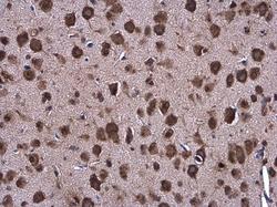
- Grp78 antibody [N2C1], Internal detects Grp78 protein at cytosol on rat hind brain by immunohistochemical analysis. Sample: Paraffin-embedded rat hind brain. Grp78 antibody [N2C1], Internal (GTX102580) dilution: 1:500.

- Submitted by
- GeneTex (provider)
- Main image
- Experimental details
- Grp78 antibody [N2C1], Internal detects Grp78 protein at cytoplasm in mouse brain by immunohistochemical analysis. Sample: Paraffin-embedded mouse brain. Grp78 antibody [N2C1], Internal (GTX102580) diluted at 1:500.

- Submitted by
- GeneTex (provider)
- Main image

- Experimental details
- Grp78 antibody [N2C1], Internal detects Grp78 protein at cytoplasm in rat prostate by immunohistochemical analysis. Sample: Paraffin-embedded rat prostate. Grp78 antibody [N2C1], Internal (GTX102580) diluted at 1:500.
