Explore
 Validate
 Learn
Learn Western blot
 Immunocytochemistry
Antibody data
- Antibody Data
- Antigen structure
- References [0]
- Comments [0]
- Validations
- Western blot [2]
- Immunocytochemistry [5]
- Immunohistochemistry [6]
Submit
Validation data
Reference
Comment
Report error
- Product number
- AMAb91010 - Provider product page

- Provider
- Atlas Antibodies
- Proper citation
- Atlas Antibodies Cat#AMAb91010, RRID:AB_2665761
- Product name
- Anti-HNRNPC
- Antibody type
- Monoclonal
- Reactivity
- Human
- Host
- Mouse
- Conjugate
- Unconjugated
- Antigen sequence
AEMYGSVTEHPSPSPLLSSS- Isotype
- IgG
- Antibody clone number
- CL2593
- Vial size
- 100 µl
- Storage
- Store at +4°C for short term storage. Long time storage is recommended at -20°C.
No comments: Submit comment
Enhanced validation
- Submitted by
- Atlas Antibodies (provider)
- Enhanced method
- Genetic validation
- Main image

- Experimental details
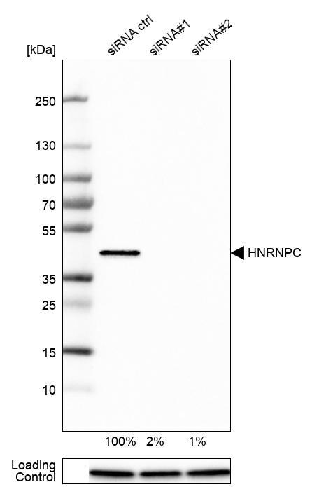
- Western blot analysis in U-251MG cells transfected with control siRNA, target specific siRNA probe #1 and #2, using Anti-HNRNPC antibody. Remaining relative intensity is presented. Loading control: Anti-PPIB.
- Submitted by
- Atlas Antibodies (provider)
- Main image

- Experimental details
- Lane 1: Marker [kDa]Lane 2:Human cell line U-251 MG

Supportive validation
- Submitted by
- Atlas Antibodies (provider)
- Main image

- Experimental details
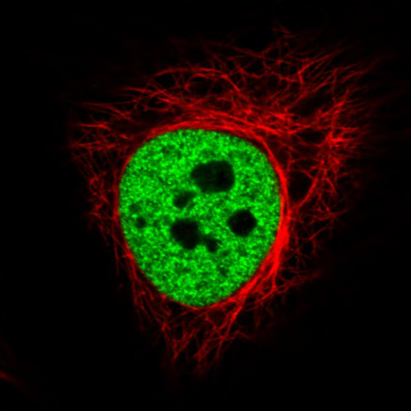
- Immunofluorescence staining in HeLa cell line with Anti-HNRNPC monoclonal antibody, showing distinct nuclear (without nucleoli) staining in green. Microtubule- and nuclear probes are visualized in red and blue respectively (where available).
- Sample type
- HUMAN
- Submitted by
- Atlas Antibodies (provider)
- Main image

- Experimental details
- Immunofluorescence staining in A431 cell line with Anti-HNRNPC monoclonal antibody, showing distinct nuclear (without nucleoli) staining in green. Microtubule- and nuclear probes are visualized in red and blue respectively (where available).
- Sample type
- HUMAN

- Submitted by
- Atlas Antibodies (provider)
- Main image

- Experimental details
- Immunofluorescence staining in MCF7 cell line with Anti-HNRNPC monoclonal antibody, showing distinct nuclear (without nucleoli) staining in green. Microtubule- and nuclear probes are visualized in red and blue respectively (where available).
- Sample type
- HUMAN

- Submitted by
- Atlas Antibodies (provider)
- Main image

- Experimental details
- Immunofluorescence staining in U2OS cell line with Anti-HNRNPC monoclonal antibody, showing distinct nuclear (without nucleoli) staining in green. Microtubule- and nuclear probes are visualized in red and blue respectively (where available).
- Sample type
- HUMAN

- Submitted by
- Atlas Antibodies (provider)
- Main image

- Experimental details
- Immunofluorescence staining in U251 cell line with Anti-HNRNPC monoclonal antibody, showing distinct nuclear (without nucleoli) staining in green. Microtubule- and nuclear probes are visualized in red and blue respectively (where available).
- Sample type
- HUMAN

Supportive validation
- Submitted by
- Atlas Antibodies (provider)
- Main image

- Experimental details
- Immunohistochemical staining of human rectum shows strong nuclear immunoreactivity in glandular and lamina propria cells.

- Submitted by
- Atlas Antibodies (provider)
- Main image
- Experimental details
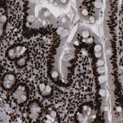
- Immunohistochemical staining of human small intestine shows strong nuclear positivity in glandular epithelium and in lamina propria cells.

- Submitted by
- Atlas Antibodies (provider)
- Main image

- Experimental details
- Immunohistochemical staining of human kidney shows strong nuclear immunoreactivity.

- Submitted by
- Atlas Antibodies (provider)
- Main image
- Experimental details
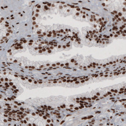
- Immunohistochemical staining of human prostate shows strong nuclear positivity in glandular and connective tissue cells.

- Submitted by
- Atlas Antibodies (provider)
- Main image

- Experimental details
- Immunohistochemical staining of human fallopian tube shows strong nuclear immunoreactivity.

- Submitted by
- Atlas Antibodies (provider)
- Main image

- Experimental details
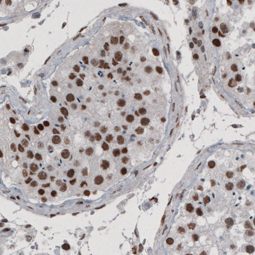
- Immunohistochemical staining of human testis shows nuclear positivity in seminiferous tubules.