 Explore
 Validate
 Learn
Learn Western blot
 Immunocytochemistry
Antibody data
- Antibody Data
- Antigen structure
- References [6]
- Comments [0]
- Validations
- Immunocytochemistry [1]
Submit
Validation data
Reference
Comment
Report error
- Product number
- MAB4060 - Provider product page

- Provider
- R&D Systems
- Product name
- Human Integrin beta 4/CD104 Antibody
- Antibody type
- Monoclonal
- Description
- Protein A or G purified from hybridoma culture supernatant. Detects human Integrin beta 4/CD104 in Western blots. In Western blots, no cross-reactivity with recombinant human Integrin beta 1, beta 2, beta 3, beta 5, beta 6, beta 8, or recombinant mouse Integrin beta 4 is observed.
- Reactivity
- Human
- Host
- Mouse
- Conjugate
- Unconjugated
- Antigen sequence
P16144- Isotype
- IgG
- Antibody clone number
- 422325
- Vial size
- 100 ug
- Concentration
- LYOPH
- Storage
- Use a manual defrost freezer and avoid repeated freeze-thaw cycles. 12 months from date of receipt, -20 to -70 °C as supplied. 1 month, 2 to 8 °C under sterile conditions after reconstitution. 6 months, -20 to -70 °C under sterile conditions after reconstitution.
Submitted references Clinical impact of different exosomes' protein expression in pancreatic ductal carcinoma patients treated with standard first line palliative chemotherapy.
Granzyme B is elevated in autoimmune blistering diseases and cleaves key anchoring proteins of the dermal-epidermal junction.
Enteric Species F Human Adenoviruses use Laminin-Binding Integrins as Co-Receptors for Infection of Ht-29 Cells.
Human Adenovirus Type 37 Uses αVβ1 and α3β1 Integrins for Infection of Human Corneal Cells.
Integrin α6β4 identifies human distal lung epithelial progenitor cells with potential as a cell-based therapy for cystic fibrosis lung disease.
Integrin α5β1 facilitates cancer cell invasion through enhanced contractile forces.
Giampieri R, Piva F, Occhipinti G, Bittoni A, Righetti A, Pagliaretta S, Murrone A, Bianchi F, Amantini C, Giulietti M, Ricci G, Principato G, Santoni G, Berardi R, Cascinu S
PloS one 2019;14(5):e0215990
PloS one 2019;14(5):e0215990
Granzyme B is elevated in autoimmune blistering diseases and cleaves key anchoring proteins of the dermal-epidermal junction.
Russo V, Klein T, Lim DJ, Solis N, Machado Y, Hiroyasu S, Nabai L, Shen Y, Zeglinski MR, Zhao H, Oram CP, Lennox PA, Van Laeken N, Carr NJ, Crawford RI, Franzke CW, Overall CM, Granville DJ
Scientific reports 2018 Jun 26;8(1):9690
Scientific reports 2018 Jun 26;8(1):9690
Enteric Species F Human Adenoviruses use Laminin-Binding Integrins as Co-Receptors for Infection of Ht-29 Cells.
Rajan A, Persson BD, Frängsmyr L, Olofsson A, Sandblad L, Heino J, Takada Y, Mould AP, Schnapp LM, Gall J, Arnberg N
Scientific reports 2018 Jul 3;8(1):10019
Scientific reports 2018 Jul 3;8(1):10019
Human Adenovirus Type 37 Uses αVβ1 and α3β1 Integrins for Infection of Human Corneal Cells.
Storm RJ, Persson BD, Skalman LN, Frängsmyr L, Lindström M, Rankin G, Lundmark R, Domellöf FP, Arnberg N
Journal of virology 2017 Mar 1;91(5)
Journal of virology 2017 Mar 1;91(5)
Integrin α6β4 identifies human distal lung epithelial progenitor cells with potential as a cell-based therapy for cystic fibrosis lung disease.
Li X, Rossen N, Sinn PL, Hornick AL, Steines BR, Karp PH, Ernst SE, Adam RJ, Moninger TO, Levasseur DN, Zabner J
PloS one 2013;8(12):e83624
PloS one 2013;8(12):e83624
Integrin α5β1 facilitates cancer cell invasion through enhanced contractile forces.
Mierke CT, Frey B, Fellner M, Herrmann M, Fabry B
Journal of cell science 2011 Feb 1;124(Pt 3):369-83
Journal of cell science 2011 Feb 1;124(Pt 3):369-83
No comments: Submit comment
Supportive validation
- Submitted by
- R&D Systems (provider)
- Main image
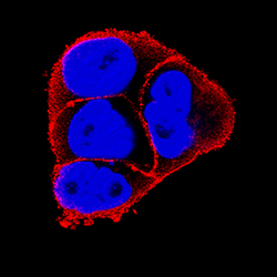
- Experimental details
- Integrin beta 4/CD104 in A431 Human Cell Line. Integrin beta 4/CD104 was detected in immersion fixed A431 human epithelial carcinoma cell line using Mouse Anti-Human Integrin beta 4/CD104 Monoclonal Antibody (Catalog # MAB4060) at 25 µg/mL for 3 hours at room temperature. Cells were stained using the NorthernLights™ 557-conjugated Anti-Mouse IgG Secondary Antibody (red; Catalog # NL007) and counterstained with DAPI (blue). Specific staining was localized to cell surfaces. View our protocol for Fluorescent ICC Staining of Cells on Coverslips.
