Explore
 Validate
 Learn
Learn Western blot
 Immunocytochemistry
Antibody data
- Antibody Data
- Antigen structure
- References [5]
- Comments [0]
- Validations
- Western blot [5]
- Immunocytochemistry [1]
- Immunoprecipitation [1]
Submit
Validation data
Reference
Comment
Report error
- Product number
- GTX100637 - Provider product page

- Provider
- GeneTex
- Proper citation
- GeneTex Cat#GTX100637, RRID:AB_1950910
- Product name
- c-Met antibody [C3], C-term
- Antibody type
- Polyclonal
- Reactivity
- Human
- Host
- Rabbit
Submitted references Silencing of MUC20 suppresses the malignant character of pancreatic ductal adenocarcinoma cells through inhibition of the HGF/MET pathway.
TOP2A induces malignant character of pancreatic cancer through activating β-catenin signaling pathway.
Loss of cell invasiveness through PKC-mediated syndecan-1 downregulation in melanoma cells under anchorage independency.
C1GALT1 enhances proliferation of hepatocellular carcinoma cells via modulating MET glycosylation and dimerization.
Translocation of Helicobacter pylori CagA into Human B lymphocytes, the origin of mucosa-associated lymphoid tissue lymphoma.
Chen ST, Kuo TC, Liao YY, Lin MC, Tien YW, Huang MC
Oncogene 2018 Nov;37(46):6041-6053
Oncogene 2018 Nov;37(46):6041-6053
TOP2A induces malignant character of pancreatic cancer through activating β-catenin signaling pathway.
Pei YF, Yin XM, Liu XQ
Biochimica et biophysica acta. Molecular basis of disease 2018 Jan;1864(1):197-207
Biochimica et biophysica acta. Molecular basis of disease 2018 Jan;1864(1):197-207
Loss of cell invasiveness through PKC-mediated syndecan-1 downregulation in melanoma cells under anchorage independency.
Wang C, Tseng T, Jhang Y, Tseng J, Hsieh C, Wu WG, Lee S
Experimental dermatology 2014 Nov;23(11):843-9
Experimental dermatology 2014 Nov;23(11):843-9
C1GALT1 enhances proliferation of hepatocellular carcinoma cells via modulating MET glycosylation and dimerization.
Wu YM, Liu CH, Huang MJ, Lai HS, Lee PH, Hu RH, Huang MC
Cancer research 2013 Sep 1;73(17):5580-90
Cancer research 2013 Sep 1;73(17):5580-90
Translocation of Helicobacter pylori CagA into Human B lymphocytes, the origin of mucosa-associated lymphoid tissue lymphoma.
Lin WC, Tsai HF, Kuo SH, Wu MS, Lin CW, Hsu PI, Cheng AL, Hsu PN
Cancer research 2010 Jul 15;70(14):5740-8
Cancer research 2010 Jul 15;70(14):5740-8
No comments: Submit comment
Enhanced validation
Supportive validation
- Submitted by
- GeneTex (provider)
- Enhanced method
- Genetic validation
- Main image
- Experimental details
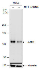
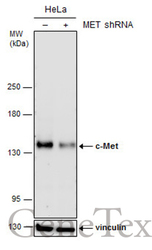
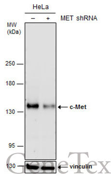
- Non-transfected (¡V) and transfected (+) HeLa whole cell extracts (30 ?g) were separated by 5% SDS-PAGE, and the membrane was blotted with c-Met antibody [C3], C-term (GTX100637) diluted at 1:500. The HRP-conjugated anti-rabbit IgG antibody (GTX213110-01) was used to detect the primary antibody.
Supportive validation
- Submitted by
- GeneTex (provider)
- Main image
- Experimental details
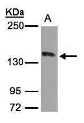
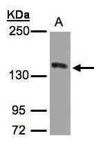
- Sample(30 ?g whole cell lysate)A:HeLa S3(GTX14654)5% SDS PAGEGTX100637 diluted at 1:1000The HRP-conjugated anti-rabbit IgG antibody (GTX213110-01) was used to detect the primary antibody.
- Submitted by
- GeneTex (provider)
- Main image
- Experimental details
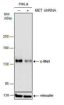
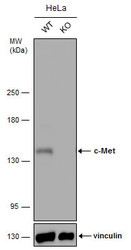
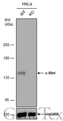
- Wild-type (WT) and c-Met knockout (KO) HeLa cell extracts (30 ?g) were separated by 5% SDS-PAGE, and the membrane was blotted with c-Met antibody [C3], C-term (GTX100637) diluted at 1:1000. The HRP-conjugated anti-rabbit IgG antibody (GTX213110-01) was used to detect the primary antibody.
- Submitted by
- GeneTex (provider)
- Main image
- Experimental details
- Non-transfected (¡V) and transfected (+) HeLa whole cell extracts (30 ?g) were separated by 5% SDS-PAGE, and the membrane was blotted with c-Met antibody [C3], C-term (GTX100637) diluted at 1:500. The HRP-conjugated anti-rabbit IgG antibody (GTX213110-01) was used to detect the primary antibody.
- Submitted by
- GeneTex (provider)
- Main image
- Experimental details
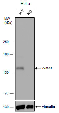
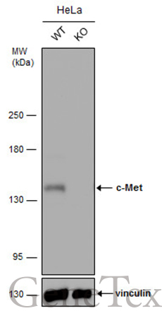
- Wild-type (WT) and c-Met knockout (KO) HeLa cell extracts (30 ?g) were separated by 5% SDS-PAGE, and the membrane was blotted with c-Met antibody [C3], C-term (GTX100637) diluted at 1:1000. The HRP-conjugated anti-rabbit IgG antibody (GTX213110-01) was used to detect the primary antibody.
Supportive validation
- Submitted by
- GeneTex (provider)
- Main image

- Experimental details
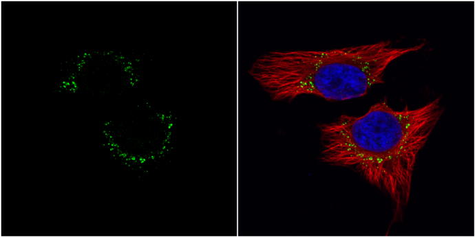
- c-Met antibody [c3], c-term detects c-Met protein at cytoplasm by immunofluorescent analysis.Sample: HeLa cells were fixed in 4% paraformaldehyde at RT for 15 min.Green: c-Met protein stained by c-Met antibody [c3], c-term (GTX100637) diluted at 1:1000.Red: alpha Tubulin, a cytoskeleton marker, stained by alpha Tubulin antibody [GT114] (GTX628802) diluted at 1:500.Blue: Hoechst 33342 staining.
Supportive validation
- Submitted by
- GeneTex (provider)
- Main image
- Experimental details
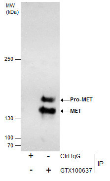
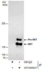
- Immunoprecipitation of c-Met protein from HeLa whole cell extracts using 5 £gg of c-Met antibody [C3], C-term (GTX100637).Western blot analysis was performed using c-Met antibody [C3], C-term (GTX100637).EasyBlot anti-Rabbit IgG (GTX221666-01) was used as a secondary reagent.