Explore
 Validate
 Learn
Learn Western blot
Antibody data
- Antibody Data
- Antigen structure
- References [0]
- Comments [0]
- Validations
- Western blot [2]
- Immunocytochemistry [2]
- Immunohistochemistry [4]
Submit
Validation data
Reference
Comment
Report error
- Product number
- HPA051280 - Provider product page

- Provider
- Atlas Antibodies
- Proper citation
- Atlas Antibodies Cat#HPA051280, RRID:AB_2681417
- Product name
- Anti-KHDRBS1
- Antibody type
- Polyclonal
- Reactivity
- Human
- Host
- Rabbit
- Conjugate
- Unconjugated
- Antigen sequence
YDDTYAEQSYEGYEGYYSQSQGDSEYYDYGHGEVQ
DSYEAYGQDDWNGTRPSLKAPPARPVKGAYR- Isotype
- IgG
- Vial size
- 100 µl
- Storage
- Store at +4°C for short term storage. Long time storage is recommended at -20°C.
No comments: Submit comment
Enhanced validation
Supportive validation
- Submitted by
- klas2
- Enhanced method
- Genetic validation
- Main image

- Experimental details
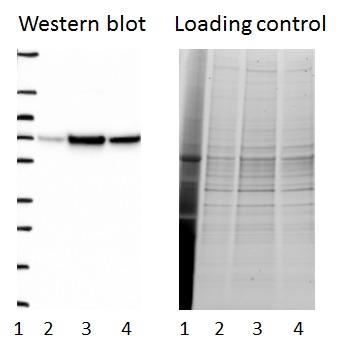
- Western blot of cell lysate from U-2 OS cells transfected with either siRNA targeting KHDRBS1 or control siRNA. Lane 1: Marker (250, 130, 95, 72, 55, 36, 28, 17, 10) Lane 2: Cell lysate from U-2OS cells transfected with siRNA targeting KHDRBS1 Lane 3: N/A Lane 4: Cell lysate from U-2OS cells transfected with control siRNA Right image, lane 1-4: loading control
- Sample type
- U-2 OS
- Primary Ab dilution
- 1:197
- Conjugate
- Horseradish Peroxidase
- Secondary Ab
- Secondary Ab
- Secondary Ab dilution
- 1:3000
- Knockdown/Genetic Approaches Application
- Western blot
Supportive validation
- Submitted by
- Atlas Antibodies (provider)
- Enhanced method
- Genetic validation
- Main image
- Experimental details
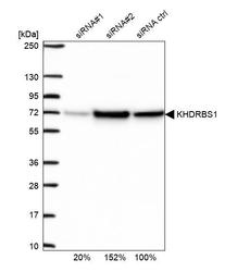
- Western blot analysis in U2OS cells transfected with control siRNA, target specific siRNA probe #1 and #2, using Anti-KHDRBS1 antibody. Remaining relative intensity is presented.

Enhanced validation
Supportive validation
- Submitted by
- 55af80e3e0991
- Enhanced method
- Genetic validation
- Main image

- Experimental details
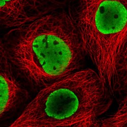
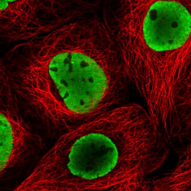
- Confocal images of immunofluorescently stained human U-2 OS cells.The protein KHDRBS1 is shown in green and the microtubules in red. The image to the left show cells transfected with control siRNA and the image to the right show cells where KHDRBS1 has been downregulated with specific siRNA.
- Sample type
- U-2 OS cells
- Primary Ab dilution
- 1:88
- Secondary Ab
- Secondary Ab
- Secondary Ab dilution
- 1:800
- Knockdown/Genetic Approaches Application
- Immunocytochemistry

Supportive validation
- Submitted by
- Atlas Antibodies (provider)
- Main image
- Experimental details
- Immunofluorescent staining of human cell line A-431 shows localization to nucleoplasm.
- Sample type
- HUMAN
Enhanced validation
Supportive validation
- Submitted by
- Atlas Antibodies (provider)
- Enhanced method
- Orthogonal validation
- Main image

- Experimental details
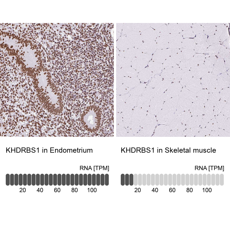
- Immunohistochemistry analysis in human endometrium and skeletal muscle tissues using Anti-KHDRBS1 antibody. Corresponding KHDRBS1 RNA-seq data are presented for the same tissues.
- Sample type
- HUMAN
Supportive validation
- Submitted by
- Atlas Antibodies (provider)
- Main image

- Experimental details
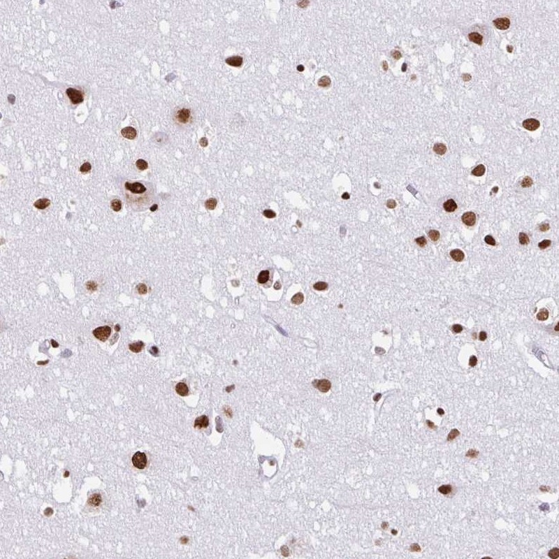
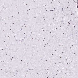
- Immunohistochemical staining of human cerebral cortex shows strong nuclear positivity in both neuronal cells and glial cells.
- Submitted by
- Atlas Antibodies (provider)
- Main image

- Experimental details
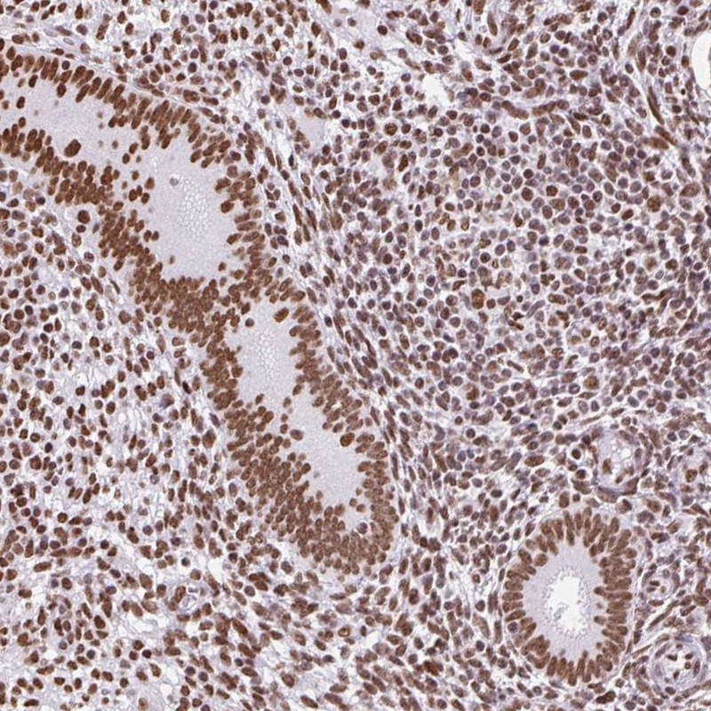
- Immunohistochemical staining of human endometrium shows high expression.
- Sample type
- HUMAN
- Submitted by
- Atlas Antibodies (provider)
- Main image
- Experimental details
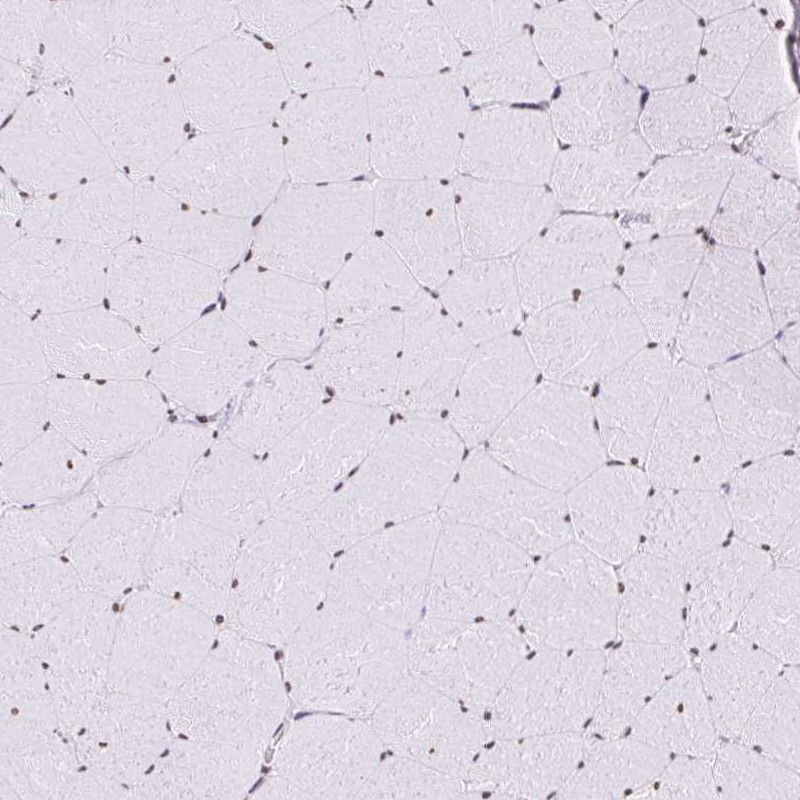
- Immunohistochemical staining of human skeletal muscle shows low expression as expected.
- Sample type
- HUMAN