Explore
 Validate
 Learn
Learn Immunocytochemistry
Antibody data
- Antibody Data
- Antigen structure
- References [0]
- Comments [0]
- Validations
- Immunocytochemistry [1]
- Immunohistochemistry [2]
Submit
Validation data
Reference
Comment
Report error
- Product number
- AF7215 - Provider product page

- Provider
- R&D Systems
- Product name
- Human/Mouse Smad9 Antibody
- Antibody type
- Polyclonal
- Description
- Immunogen affinity purified. Detects human Smad9 in direct ELISAs. In direct ELISAs, approximately 5% cross-reactivity with recombinant human Smad1 is observed.
- Reactivity
- Human, Mouse
- Host
- Sheep
- Conjugate
- Unconjugated
- Antigen sequence
O15198- Isotype
- IgG
- Vial size
- 100 ug
- Concentration
- LYOPH
- Storage
- Use a manual defrost freezer and avoid repeated freeze-thaw cycles. 12 months from date of receipt, -20 to -70 °C as supplied. 1 month, 2 to 8 °C under sterile conditions after reconstitution. 6 months, -20 to -70 °C under sterile conditions after reconstitution.
No comments: Submit comment
Supportive validation
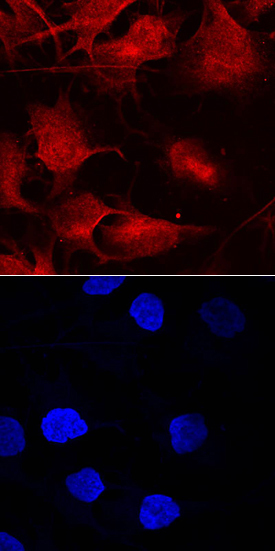
- Submitted by
- R&D Systems (provider)
- Main image

- Experimental details
- Smad9 in A172 Human Cell Line. Smad9 was detected in immersion fixed A172 human glioblastoma cell line using Sheep Anti-Human/Mouse Smad9 Antigen Affinity-purified Polyclonal Antibody (Catalog # AF7215) at 10 µg/mL for 3 hours at room temperature. Cells were stained using the NorthernLights™ 557-conjugated Anti-Sheep IgG Secondary Antibody (red, upper panel; Catalog # NL010) and counterstained with DAPI (blue, lower panel). Specific staining was localized to cytoplasm. View our protocol for Fluorescent ICC Staining of Cells on Coverslips.
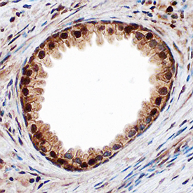
Supportive validation
- Submitted by
- R&D Systems (provider)
- Main image

- Experimental details
- Smad9 in Human Prostate Cancer Tissue. Smad9 was detected in immersion fixed paraffin-embedded sections of human prostate cancer tissue using Sheep Anti-Human Smad9 Antigen Affinity-purified Polyclonal Antibody (Catalog # AF7215) at 3 µg/mL overnight at 4 °C. Before incubation with the primary antibody, tissue was subjected to heat-induced epitope retrieval using Antigen Retrieval Reagent-Basic (Catalog # CTS013). Tissue was stained using the Anti-Sheep HRP-DAB Cell & Tissue Staining Kit (brown; Catalog # CTS019) and counterstained with hematoxylin (blue). Specific staining was localized to nuclei in glandular epithelial cells. View our protocol for Chromogenic IHC Staining of Paraffin-embedded Tissue Sections.
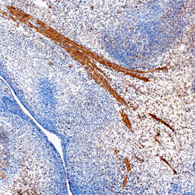
- Submitted by
- R&D Systems (provider)
- Main image

- Experimental details
- Smad9 in Mouse Embryo. Smad9 was detected in immersion fixed frozen sections of mouse embryo (13 d.p.c.) using Sheep Anti-Human Smad9 Antigen Affinity-purified Polyclonal Antibody (Catalog # AF7215) at 1.7 µg/mL overnight at 4 °C. Tissue was stained using the Anti-Sheep HRP-DAB Cell & Tissue Staining Kit (brown; Catalog # CTS019) and counterstained with hematoxylin (blue). Specific staining was localized to developing muscle cells. View our protocol for Chromogenic IHC Staining of Frozen Tissue Sections.