 Explore
 Validate
 Learn
Learn Western blot
 ELISA
ELISAAntibody data
- Antibody Data
- Antigen structure
- References [0]
- Comments [0]
- Validations
- Western blot [3]
Submit
Validation data
Reference
Comment
Report error
- Product number
- LS-C60105 - Provider product page

- Provider
- LSBio
- Proper citation
- LifeSpan Cat#LS-C60105, RRID:AB_2298844
- Product name
- NTRK3 / TRKC Antibody LS-C60105
- Antibody type
- Polyclonal
- Description
- Affinity purified
- Reactivity
- Human, Mouse, Rat
- Host
- Rabbit
- Isotype
- IgG
- Storage
- Store vial at -20°C prior to opening. This product is stable for several weeks at 4°C as an undiluted liquid. Dilute only prior to immediate use. For extended storage aliquot contents and freeze at -20°C or below. Avoid freeze-thaw cycles.
No comments: Submit comment
Supportive validation
- Submitted by
- LSBio (provider)
- Enhanced method
- Genetic validation
- Main image

- Experimental details
- Anti-TrkCT1 Antibody - Western Blot. Western blot of affinity purified anti-TrkCT1 to detect over-expressed TrkCT1 in HEK293 cells (Lane 2, arrowhead). Lane 1 is a non-transfected control. Cell extracts were resolved by electrophoresis and transferred to nitrocellulose. The membrane was probed with the primary antibody at a 1:3000 dilution. Personal Communication, V. Coppola, CCR-NCI, Frederick, MD.

- Submitted by
- LSBio (provider)
- Enhanced method
- Genetic validation
- Main image

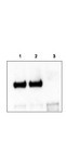
- Experimental details
- Anti-TrkCT1 Antibody - Immunoprecipitation/Western Blot. Mouse cortex lysate was immunoprecipitated with anti-TrkCT1 antibody and further blotted with affinity purified anti-TrkCT1. Lane 1 is wild-type cortex lysate, Lane 2 is Tamalin knock-out cortex lysate, and Lane 3 is TrkCT1 knock-out cortex lysate. The membrane was probed with the primary antibody at a 1:6000 dilution. Personal Communication, V. Coppola, CCR-NCI, Frederick, MD.
- Submitted by
- LSBio (provider)
- Enhanced method
- Genetic validation
- Main image

- Experimental details
- Anti-TrkCT1 Antibody - Western Blot. Western blot of affinity purified anti-TrkCT1 to detect endogenous TrkCT1 in mouse cortex lysate (Lane 1). Lane 2 is TrkCT1 knock-out cortex lysate. Cell extracts were resolved by electrophoresis and transferred to nitrocellulose. The membrane was probed with the primary antibody at a 1:6000 dilution. Personal Communication, V. Coppola, CCRNCI, Frederick, MD.
