 Explore
 Validate
 Learn
Learn Western blot
Antibody data
- Antibody Data
- Antigen structure
- References [3]
- Comments [0]
- Validations
- Western blot [2]
- Immunoprecipitation [1]
Submit
Validation data
Reference
Comment
Report error
- Product number
- PAB11334 - Provider product page

- Provider
- Abnova Corporation
- Proper citation
- Abnova Corporation Cat#PAB11334, RRID:AB_1676275
- Product name
- Ntrk3 polyclonal antibody
- Antibody type
- Polyclonal
- Description
- Rabbit polyclonal antibody raised against synthetic peptide of Ntrk3.
- Storage
- Store at 4°C. For long term storage store at -20°C.Aliquot to avoid repeated freezing and thawing.
Submitted references A kinase-deficient TrkC receptor isoform activates Arf6-Rac1 signaling through the scaffold protein tamalin.
Evidence for a role of truncated trkC receptor isoforms in mouse development.
Differential expression of TrkC catalytic and noncatalytic isoforms suggests that they act independently or in association.
Esteban PF, Yoon HY, Becker J, Dorsey SG, Caprari P, Palko ME, Coppola V, Saragovi HU, Randazzo PA, Tessarollo L
The Journal of cell biology 2006 Apr 24;173(2):291-9
The Journal of cell biology 2006 Apr 24;173(2):291-9
Evidence for a role of truncated trkC receptor isoforms in mouse development.
Palko ME, Coppola V, Tessarollo L
The Journal of neuroscience : the official journal of the Society for Neuroscience 1999 Jan 15;19(2):775-82
The Journal of neuroscience : the official journal of the Society for Neuroscience 1999 Jan 15;19(2):775-82
Differential expression of TrkC catalytic and noncatalytic isoforms suggests that they act independently or in association.
Menn B, Timsit S, Calothy G, Lamballe F
The Journal of comparative neurology 1998 Nov 9;401(1):47-64
The Journal of comparative neurology 1998 Nov 9;401(1):47-64
No comments: Submit comment
Supportive validation
- Submitted by
- Abnova Corporation (provider)
- Main image
- Experimental details
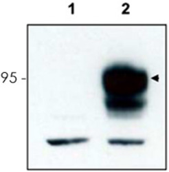
- Western blot using Ntrk3 polyclonal antibody (Cat # PAB11334) to detect over-expressed Ntrk3 in HEK293 cells (Lane 2, arrowhead). Lane 1 is a non-transfected control.Cell extracts were resolved by electrophoresis and transferred to nitrocellulose. The membrane was probed with the primary antibody at a 1:3,000 dilution.Personal Communication, V. Coppola, CCR-NCI, Frederick, MD.

- Submitted by
- Abnova Corporation (provider)
- Main image

- Experimental details
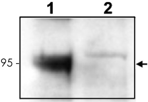
- Western blot using Ntrk3 polyclonal antibody (Cat # PAB11334) to detect endogenous Ntrk3 in mouse cortex lysate (Lane 1). Lane 2 is Ntrk3 knock-out cortex lysate.Cell extracts were resolved by electrophoresis and transferred to nitrocellulose. The membrane was probed with the primary antibody at a 1:6,000 dilution.Personal Communication, V. Coppola, CCR-NCI, Frederick, MD.
Supportive validation
- Submitted by
- Abnova Corporation (provider)
- Main image
- Experimental details
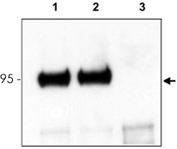
- Mouse cortex lysate was immunoprecipitated with Ntrk3 polyclonal antibody (Cat # PAB11334) and further blotted with affinity purified anti-Ntrk3.Lane 1 is wild-type cortex lysate, Lane 2 is Grasp knock-out cortex lysate, and Lane 3 is Ntrk3 knock-out cortex lysate.The membrane was probed with the primary antibody at a 1:6,000 dilution.Personal Communication, V. Coppola, CCR-NCI, Frederick, MD.
- Validation comment
- Immunoprecipitation
