 Explore
 Validate
 Learn
Learn Western blot
 ELISA
ELISA Immunoprecipitation
Antibody data
- Antibody Data
- Antigen structure
- References [0]
- Comments [0]
- Validations
- Western blot [4]
- Immunoprecipitation [1]
Submit
Validation data
Reference
Comment
Report error
- Product number
- GTX48720 - Provider product page

- Provider
- GeneTex
- Proper citation
- GeneTex Cat#GTX48720, RRID:AB_11169519
- Product name
- Trkct1 antibody
- Antibody type
- Polyclonal
- Reactivity
- Human, Mouse, Rat
- Host
- Rabbit
No comments: Submit comment
Enhanced validation
Supportive validation
- Submitted by
- GeneTex (provider)
- Enhanced method
- Genetic validation
- Main image

- Experimental details
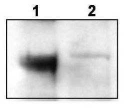
- Western blot using GeneTex's affinity purified anti-TrkCT1 to detect endogenous TrkCT1 in mouse cortex lysate (Lane 1). Lane 2 is TrkCT1 knock-out cortex lysate. Cell extracts were resolved by electrophoresis and transferred to nitrocellulose. The membrane was probed with the primary antibody at a 1:6,000 dilution.
Supportive validation
- Submitted by
- GeneTex (provider)
- Main image
- Experimental details
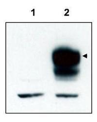
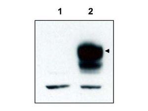
- Western blot using GeneTex's affinity purified anti-TrkCT1 to detect over-expressed TrkCT1 in HEK293 cells (Lane 2, arrowhead). Lane 1 is a non-transfected control. Cell extracts were resolved by electrophoresis and transferred to nitrocellulose. The membrane was probed with the primary antibody at a 1:3,000 dilution. Personal Communication, V. Coppola, CCR-NCI, Frederick, MD.
- Validation comment
- WB

- Submitted by
- GeneTex (provider)
- Main image

- Experimental details
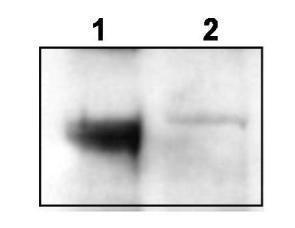
- Western blot using GeneTex's affinity purified anti-TrkCT1 to detect endogenous TrkCT1 in mouse cortex lysate (Lane 1). Lane 2 is TrkCT1 knock-out cortex lysate. Cell extracts were resolved by electrophoresis and transferred to nitrocellulose. The membrane was probed with the primary antibody at a 1:6,000 dilution. Personal Communication, V. Coppola, CCRNCI, Frederick, MD.
- Validation comment
- WB
- Submitted by
- GeneTex (provider)
- Main image
- Experimental details
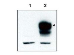
- Western blot using GeneTex's affinity purified anti-TrkCT1 to detect over-expressed TrkCT1 in HEK293 cells (Lane 2, arrowhead). Lane 1 is a non-transfected control. Cell extracts were resolved by electrophoresis and transferred to nitrocellulose. The membrane was probed with the primary antibody at a 1:3,000 dilution.
Supportive validation
- Submitted by
- GeneTex (provider)
- Main image
- Experimental details
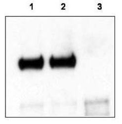
- Mouse cortex lysate was immunoprecipitated with anti-TrkCT1 antibody and further blotted with affinity purified anti-TrkCT1. Lane 1 is wild-type cortex lysate, Lane 2 is Tamalin knock-out cortex lysate, and Lane 3 is TrkCT1 knock-out cortex lysate. The membrane was probed with the primary antibody at a 1:6,000 dilution.
