Explore
 Validate
 Learn
LearnAF4984
antibody from Novus Biologicals
Targeting: IKZF1
hIk-1, Hs.54452, IKAROS, LyF-1, PPP1R92, ZNFN1A1
 Western blot
 Immunocytochemistry
Antibody data
- Antibody Data
- Antigen structure
- References [1]
- Comments [0]
- Validations
- Western blot [2]
- Flow cytometry [1]
- Chromatin Immunoprecipitation [1]
Submit
Validation data
Reference
Comment
Report error
- Product number
- AF4984 - Provider product page

- Provider
- Novus Biologicals
- Product name
- Goat Polyclonal Ikaros/IKZF1 Antibody
- Antibody type
- Polyclonal
- Description
- Antigen Affinity-purified. Detects human lkaros in direct ELISAs and Western blots. In direct ELISAs and Western blots, less than 1% cross-reactivity with recombinat human (rh) ZIC-1, rhZNF-24, and rhZNF-206 is observed.
- Reactivity
- Human
- Host
- Goat
- Conjugate
- Unconjugated
- Isotype
- IgG
- Vial size
- 100 ug
- Concentration
- LYOPH
- Storage
- Use a manual defrost freezer and avoid repeated freeze-thaw cycles. 12 months from date of receipt, -20 to -70 degreesC as supplied. 1 month, 2 to 8 degreesC under sterile conditions after reconstitution. 6 months, -20 to -70 degreesC under sterile conditions after reconstitution.
Submitted references Epstein-Barr virus utilizes Ikaros in regulating its latent-lytic switch in B cells.
Iempridee T, Reusch JA, Riching A, Johannsen EC, Dovat S, Kenney SC, Mertz JE
Journal of virology 2014 May;88(9):4811-27
Journal of virology 2014 May;88(9):4811-27
No comments: Submit comment
Supportive validation
- Submitted by
- Novus Biologicals (provider)
- Main image

- Experimental details
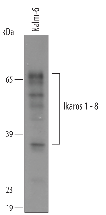
- Detection of Human Ikaros by Western Blot. Western blot shows lysates of Nalm-6 human Pre-B acute lymphocytic leukemia cell line. PVDF membrane was probed with 1 µg/mL of Goat Anti-Human Ikaros Antigen Affinity-purified Polyclonal Antibody (Catalog # AF4984) followed by HRP-conjugated Anti-Goat IgG Secondary Antibody (Catalog # HAF019). Bands were detected for Ikaros (1-8 spice forms) at approximately 37 to 63 kDa (as indicated). This experiment was conducted under reducing conditions and using Immunoblot Buffer Group 8.
- Submitted by
- Novus Biologicals (provider)
- Main image

- Experimental details
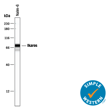
- Detection of Human Ikaros by Simple WesternTM. Simple Western lane view shows lysates of Nalm-6 human Pre-B acute lymphocytic leukemia cell line, loaded at 0.2 mg/mL. Specific bands were detected for Ikaros at approximately 63-77 kDa (as indicated) using 10 µg/mL of Goat Anti-Human Ikaros Antigen Affinity-purified Polyclonal Antibody (Catalog # AF4984) followed by 1:50 dilution of HRP-conjugated Anti-Goat IgG Secondary Antibody (Catalog # HAF109). This experiment was conducted under reducing conditions and using the 12-230 kDa separation system.
Supportive validation
- Submitted by
- Novus Biologicals (provider)
- Main image

- Experimental details
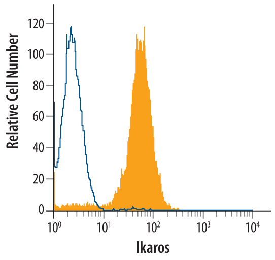
- Detection of Ikaros in Jurkat Human Cell Line by Flow Cytometry. Jurkat human acute T cell leukemia cell line was stained with Goat Anti-Human Ikaros Antigen Affinity-purified Polyclonal Antibody (Catalog # AF4984, filled histogram) or control antibody (Catalog # AB-108-C, open histogram), followed by Phycoerythrin-conjugated Anti-Goat IgG Secondary Antibody (Catalog # F0107). To facilitate intracellular staining, cells were fixed with paraformaldehyde and permeabilized with methanol.
Supportive validation
- Submitted by
- Novus Biologicals (provider)
- Main image
- Experimental details
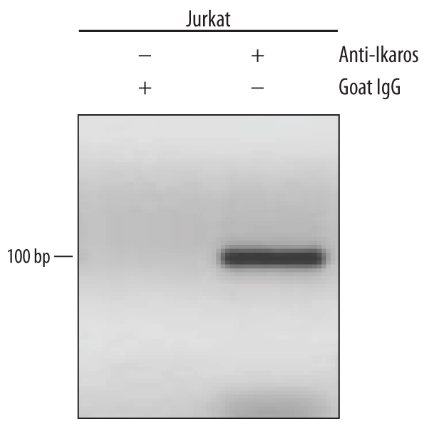
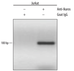
- Detection of Ikaros-regulated Genes by Chromatin Immunoprecipitation. Jurkat human acute T cell leukemia cell line treated with 50 ng/mL PMA and 200 ng/mL calcium ionomycin for 30 minutes was fixed using formaldehyde, resuspended in lysis buffer, and sonicated to shear chromatin. Ikaros/DNA complexes were immunoprecipitated using 5 μg Goat Anti-Human Ikaros Antigen Affinity-purified Polyclonal Antibody (Catalog # AF4984) or control antibody (Catalog # AB-108-C) for 15 minutes in an ultrasonic bath, followed by Biotinylated Anti-Goat IgG Secondary Antibody (Catalog # BAF109). Immunocomplexes were captured using 50 μL of MagCellect Streptavidin Ferrofluid (Catalog # MAG999) and DNA was purified using chelating resin solution. The VPAC promoter was detected by standard PCR.