Explore
 Validate
 Learn
Learn Western blot
Antibody data
- Antibody Data
- Antigen structure
- References [19]
- Comments [0]
- Validations
- Western blot [3]
- Flow cytometry [1]
- Other assay [9]
Submit
Validation data
Reference
Comment
Report error
- Product number
- PA1-020 - Provider product page

- Provider
- Invitrogen Antibodies
- Product name
- FKBP5 Polyclonal Antibody
- Antibody type
- Polyclonal
- Antigen
- Synthetic peptide
- Description
- PA1-020 detects FK506 binding protein 51 kDa (FKBP51) in canine, hamster, human, mouse and rat tissues.
- Concentration
- 1 mg/mL
Submitted references Novel Oncogenic Transcription Factor Cooperation in RB-Deficient Cancer.
Neuron-Derived Estrogen Is Critical for Astrocyte Activation and Neuroprotection of the Ischemic Brain.
Nucleocytoplasmic shuttling of the glucocorticoid receptor is influenced by tetratricopeptide repeat-containing proteins.
Heme Oxygenase 1 Impairs Glucocorticoid Receptor Activity in Prostate Cancer.
In silico identification and biochemical characterization of the human dicarboxylate clamp TPR protein interaction network.
Western High-Fat Diet Consumption during Adolescence Increases Susceptibility to Traumatic Stress while Selectively Disrupting Hippocampal and Ventricular Volumes.
Subcellular rearrangement of hsp90-binding immunophilins accompanies neuronal differentiation and neurite outgrowth.
The hsp90-FKBP52 complex links the mineralocorticoid receptor to motor proteins and persists bound to the receptor in early nuclear events.
A comparison of Hsp90alpha and Hsp90beta interactions with cochaperones and substrates.
Comparative analysis of calcineurin inhibition by complexes of immunosuppressive drugs with human FK506 binding proteins.
Ethanol-responsive brain region expression networks: implications for behavioral responses to acute ethanol in DBA/2J versus C57BL/6J mice.
Structure-function analysis of squirrel monkey FK506-binding protein 51, a potent inhibitor of glucocorticoid receptor activity.
Differential control of glucocorticoid receptor hormone-binding function by tetratricopeptide repeat (TPR) proteins and the immunosuppressive ligand FK506.
Up-regulation of glucocorticoid-regulated genes in a mouse model of Rett syndrome.
Association of immunophilins with mammalian TRPC channels.
Progesterone receptor deficient in chromatin binding has an altered cellular state.
A new first step in activation of steroid receptors: hormone-induced switching of FKBP51 and FKBP52 immunophilins.
Tissue distribution and abundance of human FKBP51, and FK506-binding protein that can mediate calcineurin inhibition.
Tissue distribution and abundance of human FKBP51, and FK506-binding protein that can mediate calcineurin inhibition.
Mandigo AC, Shafi AA, McCann JJ, Yuan W, Laufer TS, Bogdan D, Gallagher L, Dylgjeri E, Semenova G, Vasilevskaya IA, Schiewer MJ, McNair CM, de Bono JS, Knudsen KE
Cancer research 2022 Jan 15;82(2):221-234
Cancer research 2022 Jan 15;82(2):221-234
Neuron-Derived Estrogen Is Critical for Astrocyte Activation and Neuroprotection of the Ischemic Brain.
Lu Y, Sareddy GR, Wang J, Zhang Q, Tang FL, Pratap UP, Tekmal RR, Vadlamudi RK, Brann DW
The Journal of neuroscience : the official journal of the Society for Neuroscience 2020 Sep 16;40(38):7355-7374
The Journal of neuroscience : the official journal of the Society for Neuroscience 2020 Sep 16;40(38):7355-7374
Nucleocytoplasmic shuttling of the glucocorticoid receptor is influenced by tetratricopeptide repeat-containing proteins.
Mazaira GI, Echeverria PC, Galigniana MD
Journal of cell science 2020 Jun 16;133(12)
Journal of cell science 2020 Jun 16;133(12)
Heme Oxygenase 1 Impairs Glucocorticoid Receptor Activity in Prostate Cancer.
Leonardi DB, Anselmino N, Brandani JN, Jaworski FM, Páez AV, Mazaira G, Meiss RP, Nuñez M, Nemirovsky SI, Giudice J, Galigniana M, Pecci A, Gueron G, Vazquez E, Cotignola J
International journal of molecular sciences 2019 Feb 26;20(5)
International journal of molecular sciences 2019 Feb 26;20(5)
In silico identification and biochemical characterization of the human dicarboxylate clamp TPR protein interaction network.
Bernadotte A, Kumar R, Winblad B, Pavlov PF
FEBS open bio 2018 Nov;8(11):1830-1843
FEBS open bio 2018 Nov;8(11):1830-1843
Western High-Fat Diet Consumption during Adolescence Increases Susceptibility to Traumatic Stress while Selectively Disrupting Hippocampal and Ventricular Volumes.
Kalyan-Masih P, Vega-Torres JD, Miles C, Haddad E, Rainsbury S, Baghchechi M, Obenaus A, Figueroa JD
eNeuro 2016 Sep-Oct;3(5)
eNeuro 2016 Sep-Oct;3(5)
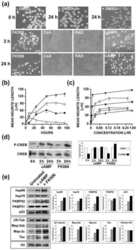
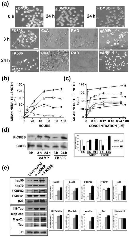
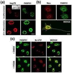
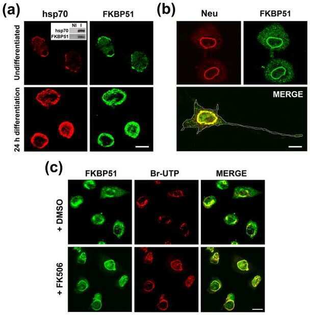
Subcellular rearrangement of hsp90-binding immunophilins accompanies neuronal differentiation and neurite outgrowth.
Quintá HR, Maschi D, Gomez-Sanchez C, Piwien-Pilipuk G, Galigniana MD
Journal of neurochemistry 2010 Nov;115(3):716-34
Journal of neurochemistry 2010 Nov;115(3):716-34
The hsp90-FKBP52 complex links the mineralocorticoid receptor to motor proteins and persists bound to the receptor in early nuclear events.
Galigniana MD, Erlejman AG, Monte M, Gomez-Sanchez C, Piwien-Pilipuk G
Molecular and cellular biology 2010 Mar;30(5):1285-98
Molecular and cellular biology 2010 Mar;30(5):1285-98
A comparison of Hsp90alpha and Hsp90beta interactions with cochaperones and substrates.
Taherian A, Krone PH, Ovsenek N
Biochemistry and cell biology = Biochimie et biologie cellulaire 2008 Feb;86(1):37-45
Biochemistry and cell biology = Biochimie et biologie cellulaire 2008 Feb;86(1):37-45
Comparative analysis of calcineurin inhibition by complexes of immunosuppressive drugs with human FK506 binding proteins.
Weiwad M, Edlich F, Kilka S, Erdmann F, Jarczowski F, Dorn M, Moutty MC, Fischer G
Biochemistry 2006 Dec 26;45(51):15776-84
Biochemistry 2006 Dec 26;45(51):15776-84
Ethanol-responsive brain region expression networks: implications for behavioral responses to acute ethanol in DBA/2J versus C57BL/6J mice.
Kerns RT, Ravindranathan A, Hassan S, Cage MP, York T, Sikela JM, Williams RW, Miles MF
The Journal of neuroscience : the official journal of the Society for Neuroscience 2005 Mar 2;25(9):2255-66
The Journal of neuroscience : the official journal of the Society for Neuroscience 2005 Mar 2;25(9):2255-66
Structure-function analysis of squirrel monkey FK506-binding protein 51, a potent inhibitor of glucocorticoid receptor activity.
Denny WB, Prapapanich V, Smith DF, Scammell JG
Endocrinology 2005 Jul;146(7):3194-201
Endocrinology 2005 Jul;146(7):3194-201
Differential control of glucocorticoid receptor hormone-binding function by tetratricopeptide repeat (TPR) proteins and the immunosuppressive ligand FK506.
Davies TH, Ning YM, Sánchez ER
Biochemistry 2005 Feb 15;44(6):2030-8
Biochemistry 2005 Feb 15;44(6):2030-8
Up-regulation of glucocorticoid-regulated genes in a mouse model of Rett syndrome.
Nuber UA, Kriaucionis S, Roloff TC, Guy J, Selfridge J, Steinhoff C, Schulz R, Lipkowitz B, Ropers HH, Holmes MC, Bird A
Human molecular genetics 2005 Aug 1;14(15):2247-56
Human molecular genetics 2005 Aug 1;14(15):2247-56
Association of immunophilins with mammalian TRPC channels.
Sinkins WG, Goel M, Estacion M, Schilling WP
The Journal of biological chemistry 2004 Aug 13;279(33):34521-9
The Journal of biological chemistry 2004 Aug 13;279(33):34521-9
Progesterone receptor deficient in chromatin binding has an altered cellular state.
Botos J, Xian W, Smith DF, Smith CL
The Journal of biological chemistry 2004 Apr 9;279(15):15231-9
The Journal of biological chemistry 2004 Apr 9;279(15):15231-9
A new first step in activation of steroid receptors: hormone-induced switching of FKBP51 and FKBP52 immunophilins.
Davies TH, Ning YM, Sánchez ER
The Journal of biological chemistry 2002 Feb 15;277(7):4597-600
The Journal of biological chemistry 2002 Feb 15;277(7):4597-600
Tissue distribution and abundance of human FKBP51, and FK506-binding protein that can mediate calcineurin inhibition.
Baughman G, Wiederrecht GJ, Chang F, Martin MM, Bourgeois S
Biochemical and biophysical research communications 1997 Mar 17;232(2):437-43
Biochemical and biophysical research communications 1997 Mar 17;232(2):437-43
No comments: Submit comment
Supportive validation
- Submitted by
- Invitrogen Antibodies (provider)
- Main image

- Experimental details
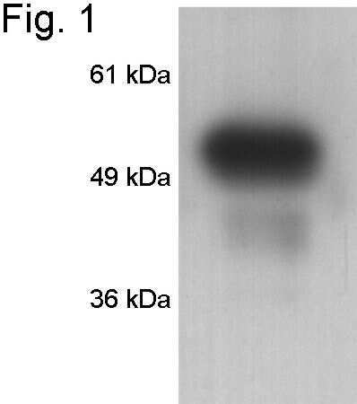
- Western blot of FKBP51 on HeLa cell lysate using Product # PA1-020.
- Submitted by
- Invitrogen Antibodies (provider)
- Main image
- Experimental details
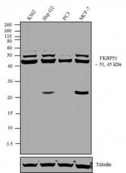
- Western blot analysis was performed on nuclear enriched extracts (30 µg lysate) of K562 (Lane 1), Hep G2 (Lane 2), PC3 (Lane 3), and MCF-7 (Lane 4). The blots were probed with Anti-FKBP51 Rabbit Polyclonal Antibody (Product # PA1-020, 1-2 µg/mL) and detected by chemiluminescence using Goat anti-Rabbit IgG (H+L) Superclonal™ Secondary Antibody, HRP conjugate (Product # A27036, 0.4 µg/mL, 1:2500 dilution). Approximately 51 and 45 kDa bands corresponding to FKBP51 were observed across cell lines tested. Known quantity of protein samples were electrophoresed using Novex® NuPAGE® 12 % Bis-Tris gel (Product # NP0342BOX), XCell SureLock™ Electrophoresis System (Product # EI0002) and Novex® Sharp Pre-Stained Protein Standard (Product # LC5800). Resolved proteins were then transferred onto a nitrocellulose membrane with iBlot® 2 Dry Blotting System (Product # IB21001). The membrane was probed with the relevant primary and secondary Antibody following blocking with 5 % skimmed milk. Chemiluminescent detection was performed using Pierce™ ECL Western Blotting Substrate (Product # 32106).

- Submitted by
- Invitrogen Antibodies (provider)
- Main image
- Experimental details
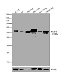
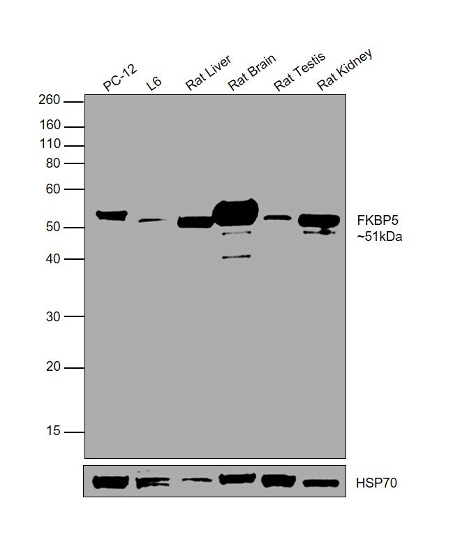
- Western blot was performed using Anti-FKBP5 Polyclonal Antibody (Product # PA1-020) and a 51kDa band corresponding to FKBP5 was observed across all the tested cell lines and tissues. Whole cell extracts (30 µg lysate) of PC-12 (Lane 1), L6 (Lane 2), Rat Liver (Lane 3), Rat Brain (Lane 4), Rat Testis (Lane 5), Rat Kidney (Lane 6) were electrophoresed using NuPAGE™ 10% Bis-Tris Protein Gel (Product # NP0301BOX). Resolved proteins were then transferred onto a Nitrocellulose membrane (Product # IB23001) by iBlot® 2 Dry Blotting System (Product # IB21001). The blot was probed with the primary antibody (1µg/mL) and detected by chemiluminescence with Goat anti-Rabbit IgG (H+L) Superclonal™ Recombinant Secondary Antibody, HRP (Product # A27036, 1:4000 dilution) using the iBright FL 1000 (Product # A32752). Chemiluminescent detection was performed using Novex® ECL Reagent Kit (Product # WP20005).
Supportive validation
- Submitted by
- Invitrogen Antibodies (provider)
- Main image
- Experimental details
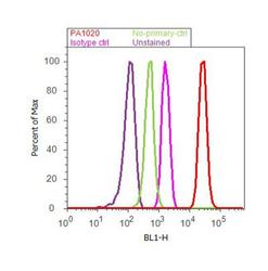
- Flow cytometry analysis of FKBP51 was done on Jurkat cells. Cells were fixed with 70% ethanol for 10 minutes, permeabilized with 0.25% Triton™ X-100 for 20 minutes, and blocked with 5% BSA for 30 minutes at room temperature. Cells were labeled with FKBP51 Rabbit Polyclonal Antibody (PA1-020, red histogram) or with rabbit isotype control (pink histogram) at 3-5 ug/million cells in 2.5% BSA. After incubation at room temperature for 2 hours, the cells were labeled with Alexa Fluor® 488 Goat Anti-Rabbit Secondary Antibody (A11008) at a dilution of 1:400 for 30 minutes at room temperature. The representative 10, 000 cells were acquired and analyzed for each sample using an Attune® Acoustic Focusing Cytometer. The purple histogram represents unstained control cells and the green histogram represents no-primary-antibody control.
Supportive validation
- Submitted by
- Invitrogen Antibodies (provider)
- Main image
- Experimental details
- NULL

- Submitted by
- Invitrogen Antibodies (provider)
- Main image
- Experimental details
- NULL
- Submitted by
- Invitrogen Antibodies (provider)
- Main image
- Experimental details
- NULL
- Submitted by
- Invitrogen Antibodies (provider)
- Main image
- Experimental details
- NULL
- Submitted by
- Invitrogen Antibodies (provider)
- Main image

- Experimental details
- NULL
- Submitted by
- Invitrogen Antibodies (provider)
- Main image
- Experimental details
- NULL

- Submitted by
- Invitrogen Antibodies (provider)
- Main image
- Experimental details
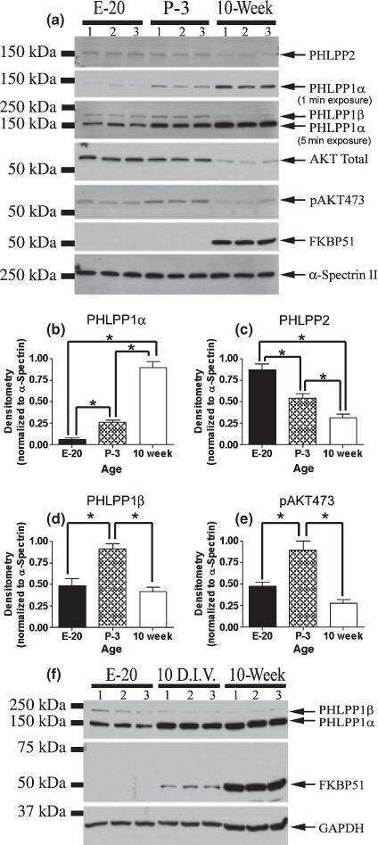
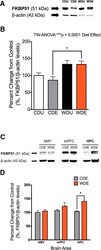
- Figure 6. WD increases FKBP51 protein levels in the hippocampus. A , Representative Western blot shows increased FKBP51 levels in the brains of the rats that consumed the WD. B , Analyses showed a significant main effect of the diet on brain FKBP51 levels (diet effect: F (1,19) = 15.45, p = 0.0009), PS exposure ( F (1,19) = 0.51, p = 0.49), or interaction between diet and PS exposure ( F (1,19) = 0.42, p = 0.52) did not show significant main effects on FKBP51 levels. C , Representative blot from micropunched anxiety-related brain regions showed distinctive FKBP51 levels. D , Analyses revealed a significant increase in FKBP51 levels in the hippocampus of the WDE rats when compared with CDE rats ( F (1,16) = 9.88; p = 0.0063, p = 0.021). Diet did not alter FKBP51 levels in the amygdala or medial prefrontal cortex. Although it did not reach statistical significance, we found a trend for more prefrontal cortex FKBP51 levels when compared with the amygdala and the hippocampus ( F (2,16) = 3.58, p = 0.052). No significant interactions between diet and the studied brain regions were revealed ( F (2,16) = 2.44, p = 0.12). Data are expressed as the mean +- SEM. * p < 0.05. Amygdala (AMY), n = 3-4 rats; medial prefrontal cortex (mPFC), n = 3-4; HPC, n = 4-5.
- Submitted by
- Invitrogen Antibodies (provider)
- Main image

- Experimental details
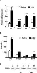
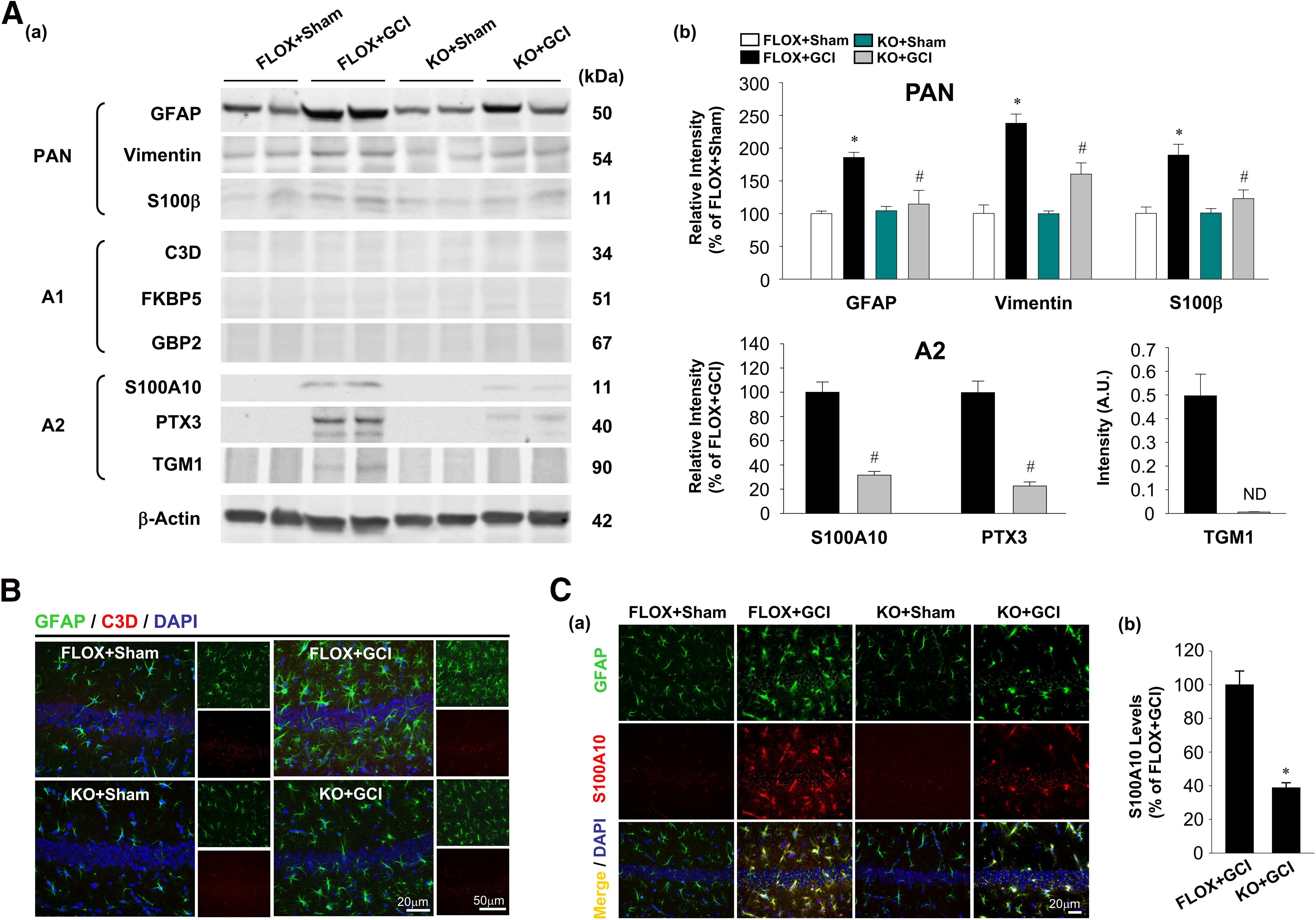
- Figure 4. Ovariectomized female FBN-ARO-KO mice have diminished astrocyte A2 phenotype 7 d after GCI. Aa , Astrocyte PAN-reactive, A1-specific, and A2-specific phenotypes were determined by Western blot analysis with purified astrocyte lysates from ovariectomized female brains 7 d after GCI reperfusion. Ab , Levels of the examined markers for different astrocyte phenotypes were quantitatively analyzed. N = 3. B , IHC analysis for astrocyte A1 phenotype by double staining of the selected astrocyte A1 marker C3D with GFAP. Ca , IHC examination for astrocyte A2 phenotype using A2-specific marker S100A10 and GFAP double staining. Cb , Percentage changes of S100A10 in FBN-ARO-KO+GCI mice versus FLOX+GCI mice were quantified. N = 4. Values are mean +- SEM of determinations from each group. * p < 0.05 versus FLOX+Sham. # p < 0.05 versus FLOX+GCI. ND, Nondetectable.
- Submitted by
- Invitrogen Antibodies (provider)
- Main image

- Experimental details
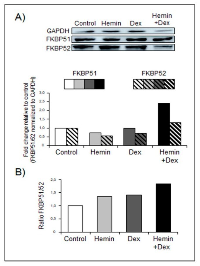
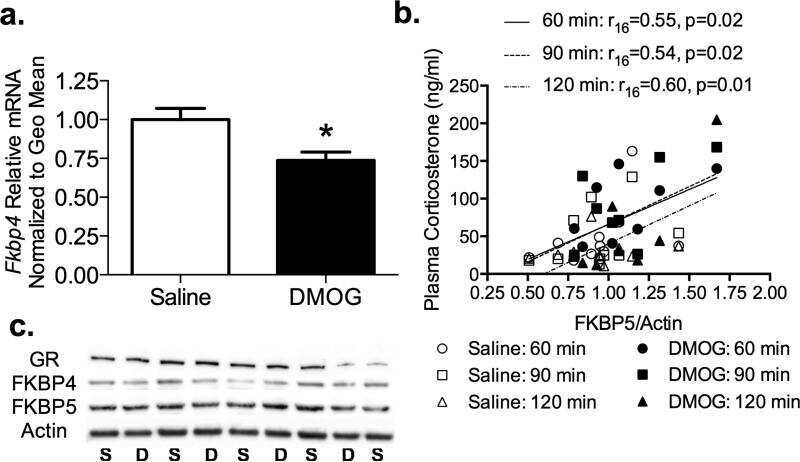
- Figure 4 Hemin increases FKBP51 expression under Dexamethasone stimulation. ( A ) Western blot analysis showing FKBP51 and FKBP52 expression in PC3 cells treated with Hemin (80 muM for 24 h), Dex (10 nM for 6 h post Hemin/PBS 24-h treatment), the combination of both drugs, or PBS as control. Total protein was extracted and protein expression was analyzed by western blotting using specific antibodies. GAPDH levels are shown as control for equal loading. Protein quantification was performed by densitometry analysis using ImageJ software and bands were normalized to GAPDH and control. ( B ) FKBP51/FKBP52 ratio was calculated for each condition. One representative from at least three independent experiments is shown.