 Explore
 Validate
 Learn
Learn Western blot
 Immunocytochemistry
Antibody data
- Antibody Data
- Antigen structure
- References [1]
- Comments [0]
- Validations
- Western blot [2]
- ELISA [1]
- Immunohistochemistry [1]
- Flow cytometry [1]
Submit
Validation data
Reference
Comment
Report error
- Product number
- NBP2-37585 - Provider product page

- Provider
- Novus Biologicals
- Product name
- Mouse Monoclonal BLNK Antibody
- Antibody type
- Monoclonal
- Description
- Unpurified.
- Reactivity
- Human, Mouse
- Host
- Mouse
- Isotype
- IgG
- Vial size
- 0.1 ml
- Storage
- Store at 4C short term. Aliquot and store at -20C long term. Avoid freeze-thaw cycles.
Submitted references Prognostic value of B-cell linker protein in colorectal cancer.
Lee JH, Lee JH, Ahn BK, Paik SS, Lee KH
Pathology, research and practice 2020 Mar;216(3):152821
Pathology, research and practice 2020 Mar;216(3):152821
No comments: Submit comment
Supportive validation
- Submitted by
- Novus Biologicals (provider)
- Main image

- Experimental details
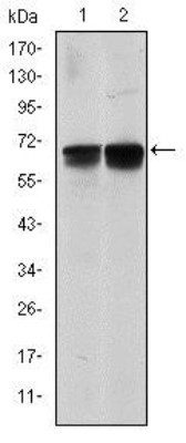
- Western Blot: BLNK Antibody (5G9) [NBP2-37585] - Western blot analysis using BLNK mouse mAb against NIH/3T3 (1) and BCBL-1 (2) cell lysate.
- Submitted by
- Novus Biologicals (provider)
- Main image

- Experimental details
- Western Blot: BLNK Antibody (5G9) [NBP2-37585] - Western blot analysis using BLNK mAb against human BLNK (AA: 34-216) recombinant protein. (Expected MW is 60 kDa)

Supportive validation
- Submitted by
- Novus Biologicals (provider)
- Main image
- Experimental details
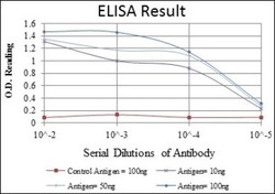
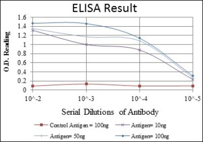
- ELISA: BLNK Antibody (5G9) [NBP2-37585] - Red: Control Antigen (100ng); Purple: Antigen (10ng); Green: Antigen (50ng); Blue: Antigen (100ng);
Supportive validation
- Submitted by
- Novus Biologicals (provider)
- Main image
- Experimental details
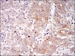
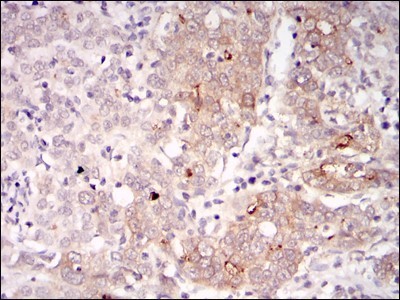
- Immunohistochemistry-Paraffin: BLNK Antibody (5G9) [NBP2-37585] - Analysis of human cervical cancer tissues using BLNK mouse mAb with DAB staining.
Supportive validation
- Submitted by
- Novus Biologicals (provider)
- Main image
- Experimental details
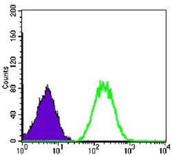
- Flow Cytometry: BLNK Antibody (5G9) [NBP2-37585] - Flow cytometric analysis of NIH/3T3 cells using BLNK mouse mAb (green) and negative control (purple).
