Explore
 Validate
 Learn
Learn Western blot
Antibody data
- Antibody Data
- Antigen structure
- References [0]
- Comments [0]
- Validations
- Western blot [2]
- Immunohistochemistry [1]
Submit
Validation data
Reference
Comment
Report error
- Product number
- AF4966 - Provider product page

- Provider
- R&D Systems
- Product name
- Human BLNK Antibody
- Antibody type
- Polyclonal
- Description
- Antigen Affinity-purified. Detects endogenous human BLNK in Western blots.
- Reactivity
- Human
- Host
- Goat
- Conjugate
- Unconjugated
- Antigen sequence
Q8WV28- Isotype
- IgG
- Vial size
- 100 ug
- Concentration
- LYOPH
- Storage
- Use a manual defrost freezer and avoid repeated freeze-thaw cycles. 12 months from date of receipt, -20 to -70 °C as supplied. 1 month, 2 to 8 °C under sterile conditions after reconstitution. 6 months, -20 to -70 °C under sterile conditions after reconstitution.
No comments: Submit comment
Supportive validation
- Submitted by
- R&D Systems (provider)
- Main image
- Experimental details
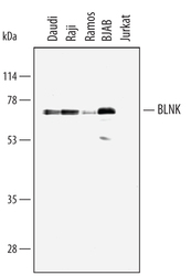
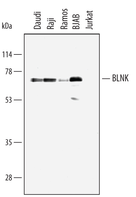
- Detection of Human BLNK by Western Blot. Western blot shows lysates of Daudi human Burkitt's lymphoma cell line, Raji human Burkitt's lymphoma cell line, Ramos human Burkitt's lymphoma cell line, BJAB human Burkitt's lymphoma cell line, and Jurkat human acute T cell leukemia cell line. PVDF membrane was probed with 1 µg/mL of Human BLNK Antigen Affinity-purified Polyclonal Antibody (Catalog # AF4966) followed by HRP-conjugated Anti-Goat IgG Secondary Antibody (Catalog # HAF109). A specific band was detected for BLNK at approximately 70 kDa (as indicated). This experiment was conducted under reducing conditions and using Immunoblot Buffer Group 1.
- Submitted by
- R&D Systems (provider)
- Main image
- Experimental details
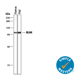
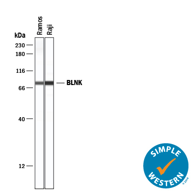
- Detection of Human BLNK by Simple WesternTM. Simple Western lane view shows lysates of Ramos human Burkitt's lymphoma cell line and Raji human Burkitt's lymphoma cell line, loaded at 0.2 mg/mL. A specific band was detected for BLNK at approximately 81 kDa (as indicated) using 10 µg/mL of Goat Anti-Human BLNK Antigen Affinity-purified Polyclonal Antibody (Catalog # AF4966) followed by 1:50 dilution of HRP-conjugated Anti-Goat IgG Secondary Antibody (Catalog # HAF109). This experiment was conducted under reducing conditions and using the 12-230 kDa separation system.
Supportive validation
- Submitted by
- R&D Systems (provider)
- Main image

- Experimental details
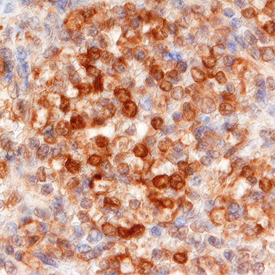
- BLNK in Human Spleen. BLNK was detected in immersion fixed paraffin-embedded sections of human spleen using Goat Anti-Human BLNK Antigen Affinity-purified Polyclonal Antibody (Catalog # AF4966) at 10 µg/mL overnight at 4 °C. Before incubation with the primary antibody, tissue was subjected to heat-induced epitope retrieval using Antigen Retrieval Reagent-Basic (Catalog # CTS013). Tissue was stained using the Anti-Goat HRP-DAB Cell & Tissue Staining Kit (brown; Catalog # CTS008) and counterstained with hematoxylin (blue). Specific staining was localized to splenocytes. View our protocol for Chromogenic IHC Staining of Paraffin-embedded Tissue Sections.