 Explore
 Validate
 Learn
Learn Western blot
Antibody data
- Antibody Data
- Antigen structure
- References [2]
- Comments [0]
- Validations
- Western blot [3]
- Immunohistochemistry [1]
Submit
Validation data
Reference
Comment
Report error
- Product number
- MAB8094 - Provider product page

- Provider
- R&D Systems
- Product name
- Human/Mouse/Rat YAP1 Antibody
- Antibody type
- Monoclonal
- Description
- Protein A or G purified from hybridoma culture supernatant. Detects human YAP1 in ELISA. Detects human, mouse and rat YAP1 in Western blots.
- Reactivity
- Human, Mouse, Rat
- Host
- Mouse
- Conjugate
- Unconjugated
- Antigen sequence
P46937- Isotype
- IgG
- Antibody clone number
- 867711
- Vial size
- 100 ug
- Concentration
- LYOPH
- Storage
- Use a manual defrost freezer and avoid repeated freeze-thaw cycles. 12 months from date of receipt, -20 to -70 °C as supplied. 1 month, 2 to 8 °C under sterile conditions after reconstitution. 6 months, -20 to -70 °C under sterile conditions after reconstitution.
Submitted references Antibody-drug conjugate T-DM1 treatment for HER2+ breast cancer induces ROR1 and confers resistance through activation of Hippo transcriptional coactivator YAP1.
A connexin43/YAP axis regulates astroglial-mesenchymal transition in hemoglobin induced astrocyte activation.
Islam SS, Uddin M, Noman ASM, Akter H, Dity NJ, Basiruzzman M, Uddin F, Ahsan J, Annoor S, Alaiya AA, Al-Alwan M, Yeger H, Farhat WA
EBioMedicine 2019 May;43:211-224
EBioMedicine 2019 May;43:211-224
A connexin43/YAP axis regulates astroglial-mesenchymal transition in hemoglobin induced astrocyte activation.
Yang Y, Ren J, Sun Y, Xue Y, Zhang Z, Gong A, Wang B, Zhong Z, Cui Z, Xi Z, Yang GY, Sun Q, Bian L
Cell death and differentiation 2018 Oct;25(10):1870-1884
Cell death and differentiation 2018 Oct;25(10):1870-1884
No comments: Submit comment
Supportive validation
- Submitted by
- R&D Systems (provider)
- Main image
- Experimental details
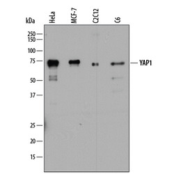
- Detection of Human, Mouse, and Rat YAP1 by Western Blot. Western blot shows lysates of HeLa human cervical epithelial carcinoma cell line, MCF-7 human breast cancer cell line, C2C12 mouse myoblast cell line, and C6 rat glioma cell line. PVDF membrane was probed with 2 µg/mL of Mouse Anti-Human YAP1 Monoclonal Antibody (Catalog # MAB8094) followed by HRP-conjugated Anti-Mouse IgG Secondary Antibody (Catalog # HAF018). A specific band was detected for YAP1 at approximately 70-75 kDa (as indicated). This experiment was conducted under reducing conditions and using Immunoblot Buffer Group 1.

- Submitted by
- R&D Systems (provider)
- Main image
- Experimental details
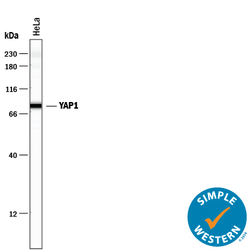
- Detection of Human YAP1 by Simple WesternTM. Simple Western lane view shows lysates of HeLa human cervical epithelial carcinoma cell line, loaded at 0.5 mg/mL. A specific band was detected for YAP1 at approximately 79 kDa (as indicated) using 20 µg/mL of Mouse Anti-Human/Mouse/Rat YAP1 Monoclonal Antibody (Catalog # MAB8094) . This experiment was conducted under reducing conditions and using the 12-230 kDa separation system.

- Submitted by
- R&D Systems (provider)
- Main image

- Experimental details
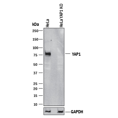
- Western Blot Shows Human YAP1 Specificity by Using Knockout Cell Line. Western blot shows lysates of HeLa human cervical epithelial carcinoma parental cell line and YAP1 knockout HeLa cell line (KO). PVDF membrane was probed with 2 µg/mL of Mouse Anti-Human/Mouse/Rat YAP1 Monoclonal Antibody (Catalog # MAB8094) followed by HRP-conjugated Anti-Mouse IgG Secondary Antibody (Catalog # HAF018). A specific band was detected for YAP1 at approximately 75 kDa (as indicated) in the parental HeLa cell line, but is not detectable in knockout HeLa cell line. GAPDH (Catalog # MAB5718) is shown as a loading control. This experiment was conducted under reducing conditions and using Immunoblot Buffer Group 1.
Supportive validation
- Submitted by
- R&D Systems (provider)
- Main image
- Experimental details
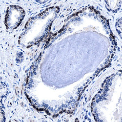
- YAP1 in Human Prostate. YAP1 was detected in formalin fixed paraffin-embedded sections of human prostate using Mouse Anti-Human YAP1 Monoclonal Antibody (Catalog # MAB8094) at 15 µg/mL overnight at 4 °C. Before incubation with the primary antibody, tissue was subjected to heat-induced epitope retrieval using Antigen Retrieval Reagent-Basic (Catalog # CTS013). Tissue was stained using the Anti-Mouse HRP-DAB Cell & Tissue Staining Kit (brown; Catalog # CTS002) and counterstained with hematoxylin (blue). Specific staining was localized to the nuclei of smooth muscle cells. View our protocol for Chromogenic IHC Staining of Paraffin-embedded Tissue Sections.
