Explore
 Validate
 Learn
Learn Western blot
 Immunocytochemistry
Antibody data
- Antibody Data
- Antigen structure
- References [2]
- Comments [0]
- Validations
- Western blot [3]
- Immunocytochemistry [1]
- Immunoprecipitation [1]
- Immunohistochemistry [1]
Submit
Validation data
Reference
Comment
Report error
- Product number
- GTX129151 - Provider product page

- Provider
- GeneTex
- Product name
- YAP1 antibody
- Antibody type
- Polyclonal
- Reactivity
- Human
- Host
- Rabbit
Submitted references Association of nuclear localization of SHP2 and YAP1 with unfavorable prognosis in non-small cell lung cancer.
Immunohistochemical studies and fluorodeoxyglucose uptake on positron emission tomography in pharyngeal cancer for predicting radiotherapy-based treatment outcomes.
Chen MJ, Wang YC, Wu DW, Chen CY, Lee H
Pathology, research and practice 2019 Apr;215(4):801-806
Pathology, research and practice 2019 Apr;215(4):801-806
Immunohistochemical studies and fluorodeoxyglucose uptake on positron emission tomography in pharyngeal cancer for predicting radiotherapy-based treatment outcomes.
Lin YC, Chen RY, Chen SW, Hsieh TC, Yen KY, Liang JA, Yang SN, Wang YC, Chen YH, Chow NH, Kao CH
Clinical otolaryngology : official journal of ENT-UK ; official journal of Netherlands Society for Oto-Rhino-Laryngology & Cervico-Facial Surgery 2017 Jun;42(3):608-619
Clinical otolaryngology : official journal of ENT-UK ; official journal of Netherlands Society for Oto-Rhino-Laryngology & Cervico-Facial Surgery 2017 Jun;42(3):608-619
No comments: Submit comment
Enhanced validation
Supportive validation
- Submitted by
- GeneTex (provider)
- Enhanced method
- Genetic validation
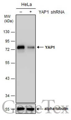
- Main image
- Experimental details
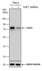
- Non-transfected (¡V) and transfected (+) HeLa whole cell extracts (30 ?g) were separated by 10% SDS-PAGE, and the membrane was blotted with YAP1 antibody (GTX129151) diluted at 1:5000. The HRP-conjugated anti-rabbit IgG antibody (GTX213110-01) was used to detect the primary antibody.

Supportive validation
- Submitted by
- GeneTex (provider)
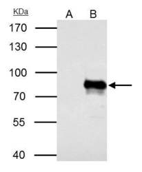
- Main image
- Experimental details
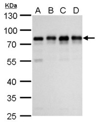
- YAP1 antibody detects YAP1 protein by western blot analysis.A. 30 ?g 293T whole cell lysate/extract B. 30 ?g A431 whole cell lysate/extract C. 30 ?g HeLa whole cell lysate/extract D. 30 ?g HepG2 whole cell lysate/extract 10% SDS-PAGEYAP1 antibody (GTX129151) dilution: 1:1000 The HRP-conjugated anti-rabbit IgG antibody (GTX213110-01) was used to detect the primary antibody.

- Submitted by
- GeneTex (provider)
- Main image
- Experimental details
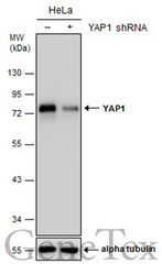
- Non-transfected (¡V) and transfected (+) HeLa whole cell extracts (30 ?g) were separated by 10% SDS-PAGE, and the membrane was blotted with YAP1 antibody (GTX129151) diluted at 1:5000. The HRP-conjugated anti-rabbit IgG antibody (GTX213110-01) was used to detect the primary antibody.
Supportive validation
- Submitted by
- GeneTex (provider)
- Main image
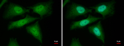
- Experimental details
- YAP1 antibody detects YAP1 protein at cytoplasm and nucleus by immunofluorescent analysis.Sample: HeLa cells were fixed in 4% paraformaldehyde at RT for 15 min.Green: YAP1 protein stained by YAP1 antibody (GTX129151) diluted at 1:1000.Blue: Hoechst 33342 staining.

Supportive validation
- Submitted by
- GeneTex (provider)
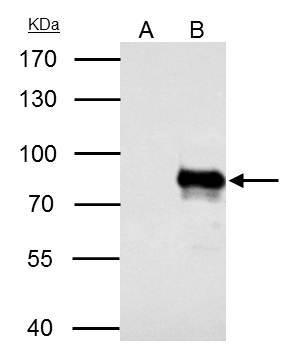
- Main image
- Experimental details
- YAP1 antibody immunoprecipitates YAP1 protein in IP experiments.IP samples: HeLa whole cell extractA. Control with 4 £gg of preimmune Rabbit IgGB. Immunoprecipitation of YAP1 protein by 4 £gg YAP1 antibody (GTX129151)7.5 % SDS-PAGEThe immunoprecipitated YAP1 protein was detected by YAP1 antibody (GTX129151) diluted at 1:500.[EasyBlot anti-rabbit IgG (GTX221666-01) was used as a secondary reagent]
Supportive validation
- Submitted by
- GeneTex (provider)
- Main image
- Experimental details
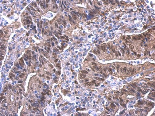
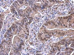
- YAP1 antibody detects YAP1 protein at nucleus on human colon carcinoma by immunohistochemical analysis. Sample: Paraffin-embedded colon carcinoma. YAP1 antibody (GTX129151) dilution: 1:500.