Explore
 Validate
 Learn
Learn Western blot
Antibody data
- Antibody Data
- Antigen structure
- References [1]
- Comments [0]
- Validations
- Western blot [3]
- Immunocytochemistry [4]
- Immunohistochemistry [3]
- Flow cytometry [1]
- Chromatin Immunoprecipitation [1]
- Other assay [1]
Submit
Validation data
Reference
Comment
Report error
- Product number
- MA5-32117 - Provider product page

- Provider
- Invitrogen Antibodies
- Product name
- YAP1 Recombinant Rabbit Monoclonal Antibody (SU33-06)
- Antibody type
- Monoclonal
- Antigen
- Synthetic peptide
- Description
- Recombinant rabbit monoclonal antibodies are produced using in vitro expression systems. The expression systems are developed by cloning in the specific antibody DNA sequences from immunoreactive rabbits. Then, individual clones are screened to select the best candidates for production. The advantages of using recombinant rabbit monoclonal antibodies include: better specificity and sensitivity, lot-to-lot consistency, animal origin-free formulations, and broader immunoreactivity to diverse targets due to larger rabbit immune repertoire.
- Reactivity
- Human
- Host
- Rabbit
- Isotype
- IgG
- Antibody clone number
- SU33-06
- Vial size
- 100 µL
- Concentration
- 1 mg/mL
- Storage
- Store at 4°C short term. For long term storage, store at -20°C, avoiding freeze/thaw cycles.
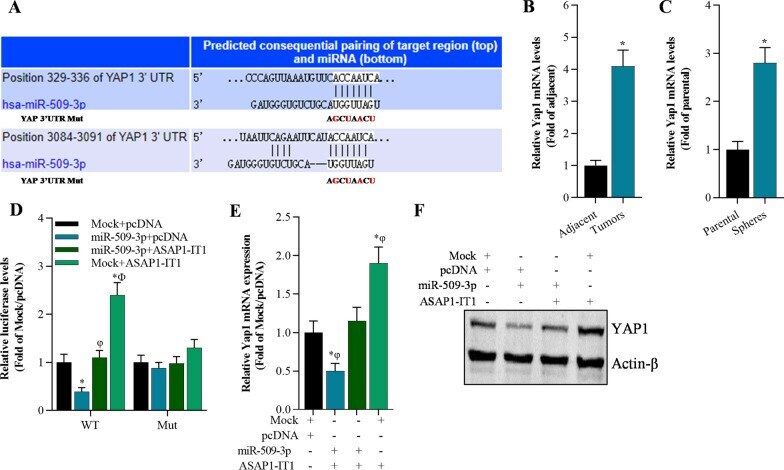
Submitted references LncRNA ASAP1-IT1 enhances cancer cell stemness via regulating miR-509-3p/YAP1 axis in NSCLC.
Liu Y, Yang Y, Zhang L, Lin J, Li B, Yang M, Li H, Chen K, Zhao W
Cancer cell international 2021 Oct 29;21(1):572
Cancer cell international 2021 Oct 29;21(1):572
No comments: Submit comment
Supportive validation
- Submitted by
- Invitrogen Antibodies (provider)
- Main image
- Experimental details
- Knockout of YAP1 was achieved by CRISPR-Cas9 genome editing using LentiArray™ Lentiviral sgRNA (Product # A32042, Assay ID CRISPR772157_LV) and LentiArray Cas9 Lentivirus (Product # A32064). Western blot analysis of YAP1 was performed by loading 30 µg of HeLa Wild Type (Lane 1), HeLa Cas9 (Lane 2) andHeLa YAP1 KO (Lane 3) whole cell extracts. The samples were electrophoresed using NuPAGE™ Novex™ 4-12% Bis-Tris Protein Gel (Product # NP0322BOX). Resolved proteins were then transferred onto a nitrocellulose membrane (Product # IB23001) by iBlot® 2 Dry Blotting System (Product # IB21001). The blot was probed with Anti-YAP1 Recombinant Rabbit Monoclonal Antibody (SU33-06) (Product # MA5-32117, 1:1,000 dilution) and Goat anti-Rabbit IgG (H+L) Superclonal™ Recombinant Secondary Antibody, HRP (Product # A27036, 1:5,000 dilution) using the iBright FL 1000 (Product # A32752). Chemiluminescent detection was performed using Novex® ECL Chemiluminescent Substrate Reagent Kit (Product # WP20005). Loss of signal upon CRISPR mediated knockout (KO) using the LentiArray™ CRISPR product line confirms that antibody is specific to YAP1.

- Submitted by
- Invitrogen Antibodies (provider)
- Main image

- Experimental details
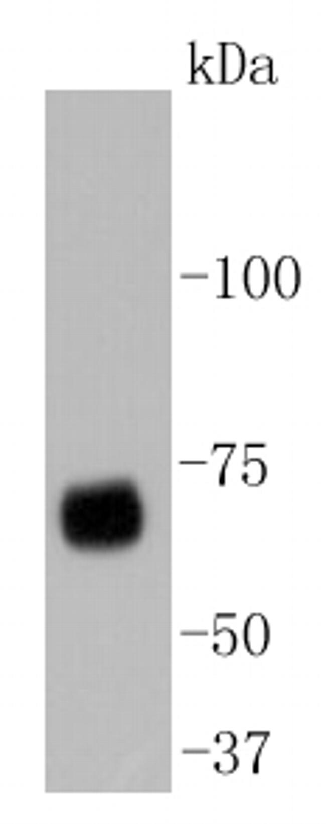
- Western blot analysis of YAP1 in HepG2 cell lysates using a YAP1 Monoclonal antibody (Product # MA5-32117) at a dilution of 1:1,000.
- Submitted by
- Invitrogen Antibodies (provider)
- Main image

- Experimental details
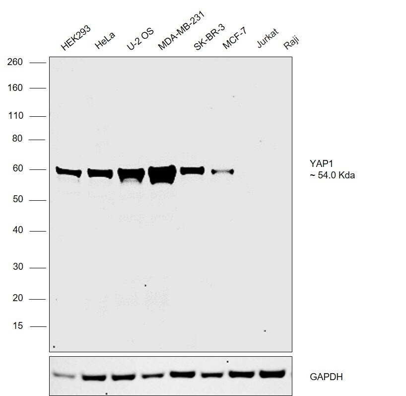
- Western blot analysis was performed on whole cell extracts (30 µg lysate) of HEK293 (Lane 1), HeLa (Lane 2), U-2 OS (Lane 3), MDA-MB-231 (Lane 4), SK-BR-3 (Lane 5), MCF-7 (Lane 6), Jurkat (Lane 7) and Raji (Lane 8). A 54 kDa band corresponding to YAP1 was observed at slightly higher molecular weight across all the cell lines tested except Jurkat and Raji which are reported to be negative. Resolved proteins were then transferred onto a nitrocellulose membrane (Product # IB23001) by iBlot® 2 Dry Blotting System (Product # IB21001) and the blot was probed with Anti-YAP1 Monoclonal Antibody (Product # MA5-32117, 1:1000 dilution) and detected by chemiluminescence with Goat anti-Rabbit IgG (H+L) Superclonal™ Recombinant Secondary Antibody, HRP conjugate (Product # A27036, 0.25 µg/ml, 1:4000 dilution) using the iBright FL 1000 (Product # A32752). Chemiluminescent detection was performed using Novex® ECL Chemiluminescent Substrate Reagent Kit (Product # WP20005).
Supportive validation
- Submitted by
- Invitrogen Antibodies (provider)
- Main image
- Experimental details
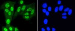
- Immunocytochemical analysis of YAP1 in Hela cells using a YAP1 Monoclonal antibody (Product # MA5-32117) as seen in green. The nuclear counter stain is DAPI (blue). Cells were fixed in paraformaldehyde, permeabilised with 0.25% Triton X100/PBS.

- Submitted by
- Invitrogen Antibodies (provider)
- Main image

- Experimental details
- Immunocytochemical analysis of YAP1 in Hela cells using a YAP1 Monoclonal antibody (Product # MA5-32117) as seen in green. The nuclear counter stain is DAPI (blue). Cells were fixed in paraformaldehyde, permeabilised with 0.25% Triton X100/PBS.

- Submitted by
- Invitrogen Antibodies (provider)
- Main image
- Experimental details
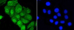
- Immunocytochemical analysis of YAP1 in HepG2 cells using a YAP1 Monoclonal antibody (Product # MA5-32117) as seen in green. The nuclear counter stain is DAPI (blue). Cells were fixed in paraformaldehyde, permeabilised with 0.25% Triton X100/PBS.

- Submitted by
- Invitrogen Antibodies (provider)
- Main image

- Experimental details
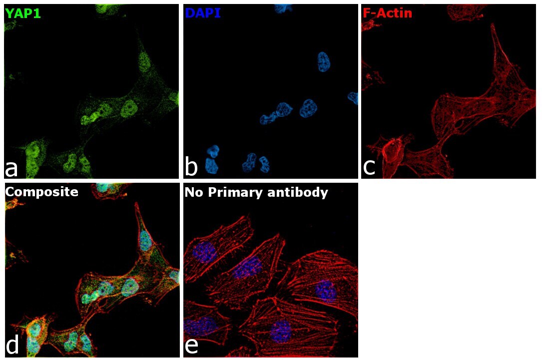
- Immunofluorescence analysis of YAP1 was performed using 70% confluent log phase MDA-MB-231 cells. The cells were fixed with 4% paraformaldehyde for 10 minutes, permeabilized with 0.1% Triton™ X-100 for 15 minutes, and blocked with 2% BSA for 1 hour at room temperature. The cells were labeled with YAP1 Recombinant Rabbit Monoclonal Antibody (Product # MA5-23117) at 5 µg/mL in 0.1% BSA, incubated at 4 degree Celsius overnight and then labeled with Goat anti-Rabbit IgG (H+L) Superclonal™ Recombinant Secondary Antibody, Alexa Fluor® 488 conjugate (Product # A27034) at a dilution of 1:2000 for 45 minutes at room temperature (Panel a: green). Nuclei (Panel b: blue) were stained with ProLong™ Diamond Antifade Mountant with DAPI (Product # P36962). F-actin (Panel c: red) was stained with Rhodamine Phalloidin (Product # R415). Panel d represents the merged image showing Nucleus and Cytoplasm localization. Panel e represents control cells with no primary antibody to assess background. The images were captured at 60X magnification.
Supportive validation
- Submitted by
- Invitrogen Antibodies (provider)
- Main image
- Experimental details
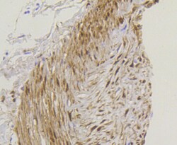
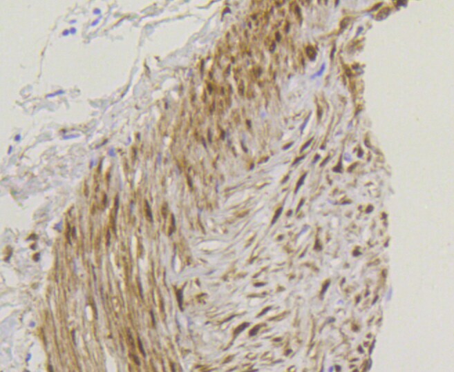
- Immunohistochemical analysis of YAP1 of paraffin-embedded Human colon cancer tissue using a YAP1 Monoclonal antibody (Product #MA5-32117). Counter stained with hematoxylin.
- Submitted by
- Invitrogen Antibodies (provider)
- Main image

- Experimental details
- Immunohistochemical analysis of YAP1 of paraffin-embedded Human breast carcinoma tissue using a YAP1 Monoclonal antibody (Product #MA5-32117). Counter stained with hematoxylin.

- Submitted by
- Invitrogen Antibodies (provider)
- Main image
- Experimental details
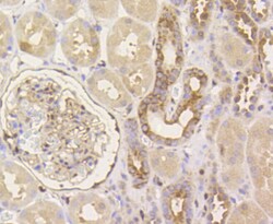
- Immunohistochemical analysis of YAP1 of paraffin-embedded Human kidney tissue using a YAP1 Monoclonal antibody (Product #MA5-32117). Counter stained with hematoxylin.

Supportive validation
- Submitted by
- Invitrogen Antibodies (provider)
- Main image

- Experimental details
- Flow Cytometric analysis of YAP1 in NIH/3T3 cells using a YAP1 Monoclonal Antibody (Product # MA5-32117) at a dilution of 1:50, as seen in blue compared with an unlabelled control (cells without incubation with primary antibody; red). Alexa Fluor 488-conjugated goat anti rabbit IgG was used as the secondary antibody.

Supportive validation
- Submitted by
- Invitrogen Antibodies (provider)
- Main image
- Experimental details
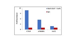
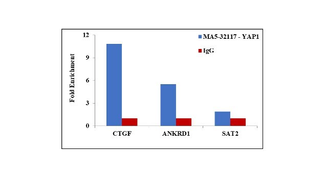
- Chromatin Immunoprecipitation (ChIP) assay of endogenous YAP1 protein using Anti-YAP1 Antibody: ChIP was performed using YAP1 Recombinant Rabbit Monoclonal Antibody (SU33-06) (Product # MA5-32117, 2.5 µg) on sheared chromatin from MDA-MB-231 cells using the MAGnify ChIP System kit (Product # 49-2024). Normal Rabbit IgG was used as a negative IP control. The purified DNA was analyzed by qPCR using primers binding to ANKRD1 and CTGF transcriptional start site and SAT2 satellite repeats. Data is presented as fold enrichment of the antibody signal versus the negative control IgG using the comparative CT method.
Supportive validation
- Submitted by
- Invitrogen Antibodies (provider)
- Main image
- Experimental details
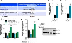
- Fig. 6 YAP1 is regulated by miR-509-3p and ASAP1-IT1. A The potential target of miR-509-3p is predicted by TargetScan. B qRT-PCR analysis of YAP1 expression in NSCLC tissues and adjacent tissues, * p < 0.01 compared with adjacent tissues. C qRT-PCR analysis of YAP1 in A549 spheres, * p < 0.01 compared with parental group. D Luciferase activity of pmirGLO-YAP1-3'UTR-WT (wild type) or pmirGLO-YAP1-3'UTR-Mut (mutant) was modulated by miR-509-3p and ASAP1-IT1 in A549 cells, * p < 0.01 compared with Mock + pcDNA, phi p < 0.01 compared with miR-509-3p + pcDNA, F p < 0.01 compared with miR-509-3p + ASAP1-IT1. E YAP1 mRNA levels in A549 cells analyzed byqRT-PCR, * p < 0.01 compared with Mock + pcDNA, phi p < 0.01 compared with miR-509-3p + pcDNA, F p < 0.01 compared with miR-509-3p + ASAP1-IT1. F Western blot analysis on YAP1 protein in A549 cells