 Explore
 Validate
 Learn
Learn Western blot
 ELISA
ELISAAntibody data
- Antibody Data
- Antigen structure
- References [6]
- Comments [0]
- Validations
- Western blot [2]
Submit
Validation data
Reference
Comment
Report error
- Product number
- GTX27523 - Provider product page

- Provider
- GeneTex
- Proper citation
- GeneTex Cat#GTX27523, RRID:AB_374462
- Product name
- Gli1 antibody
- Antibody type
- Polyclonal
- Antigen
- Other
- Description
- Antiserum
- Reactivity
- Human, Mouse
- Host
- Rabbit
- Storage
- Store vial at -20° C prior to opening. Aliquot contents and freeze at -20° C or below for extended storage. Avoid cycles of freezing and thawing.
Submitted references Identification of Human Cutaneous Basal Cell Carcinoma Cancer Stem Cells.
Comparative multidimensional molecular analyses of pediatric diffuse intrinsic pontine glioma reveals distinct molecular subtypes.
CD200-expressing human basal cell carcinoma cells initiate tumor growth.
Inhibition of hedgehog signalling prevents experimental fibrosis and induces regression of established fibrosis.
A GLI1-p53 inhibitory loop controls neural stem cell and tumour cell numbers.
Sonic hedgehog signaling pathway is activated in ALK-positive anaplastic large cell lymphoma.
Morgan H, Olivero C, Patel GK
Methods in molecular biology (Clifton, N.J.) 2019;1879:435-450
Methods in molecular biology (Clifton, N.J.) 2019;1879:435-450
Comparative multidimensional molecular analyses of pediatric diffuse intrinsic pontine glioma reveals distinct molecular subtypes.
Saratsis AM, Kambhampati M, Snyder K, Yadavilli S, Devaney JM, Harmon B, Hall J, Raabe EH, An P, Weingart M, Rood BR, Magge SN, MacDonald TJ, Packer RJ, Nazarian J
Acta neuropathologica 2014;127(6):881-95
Acta neuropathologica 2014;127(6):881-95
CD200-expressing human basal cell carcinoma cells initiate tumor growth.
Colmont CS, Benketah A, Reed SH, Hawk NV, Telford WG, Ohyama M, Udey MC, Yee CL, Vogel JC, Patel GK
Proceedings of the National Academy of Sciences of the United States of America 2013 Jan 22;110(4):1434-9
Proceedings of the National Academy of Sciences of the United States of America 2013 Jan 22;110(4):1434-9
Inhibition of hedgehog signalling prevents experimental fibrosis and induces regression of established fibrosis.
Horn A, Kireva T, Palumbo-Zerr K, Dees C, Tomcik M, Cordazzo C, Zerr P, Akhmetshina A, Ruat M, Distler O, Beyer C, Schett G, Distler JH
Annals of the rheumatic diseases 2012 May;71(5):785-9
Annals of the rheumatic diseases 2012 May;71(5):785-9
A GLI1-p53 inhibitory loop controls neural stem cell and tumour cell numbers.
Stecca B, Ruiz i Altaba A
The EMBO journal 2009 Mar 18;28(6):663-76
The EMBO journal 2009 Mar 18;28(6):663-76
Sonic hedgehog signaling pathway is activated in ALK-positive anaplastic large cell lymphoma.
Singh RR, Cho-Vega JH, Davuluri Y, Ma S, Kasbidi F, Milito C, Lennon PA, Drakos E, Medeiros LJ, Luthra R, Vega F
Cancer research 2009 Mar 15;69(6):2550-8
Cancer research 2009 Mar 15;69(6):2550-8
No comments: Submit comment
Supportive validation
- Submitted by
- GeneTex (provider)
- Main image

- Experimental details
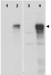
- Western blot using GeneTex anti-Gli-1 antibody (GTX27523) shows detection of a band at ~150 kDa (arrowhead) corresponding to human Gli-1 present in transfected 293T cell lysates (lanes 2 and 4). Mock 293T cell lysates with vector only show no staining (lanes 1 and 3). Lysates were separated by SDS-PAGE and transferred to nitrocellulose. After blocking the membrane was probed with the primary antibody diluted to 1:8,000 (lanes 1 and 2) or 1:4,000 (lanes 3 and 4). Molecular weight estimation was made by comparison to MW markers. Personal communication, Hiro Kimura, St. Jude Children's Research Hospital, Memphis, TN.
- Validation comment
- WB

- Submitted by
- GeneTex (provider)
- Main image
- Experimental details
- Western blot using GeneTex anti-Gli-1 antibody (GTX27523) shows detection of a band at ~150 kDa (arrowhead) corresponding to human Gli-1 present in transfected 293T cell lysates (lanes 2 and 4). Mock 293T cell lysates with vector only show no staining (lanes 1 and 3). Lysates were separated by SDS-PAGE and transferred to nitrocellulose. After blocking the membrane was probed with the primary antibody diluted to 1:8,000 (lanes 1 and 2) or 1:4,000 (lanes 3 and 4). Molecular weight estimation was made by comparison to MW markers. Personal communication, Hiro Kimura, St. Jude Children's Research Hospital, Memphis, TN.
- Validation comment
- WB
