Explore
 Validate
 Learn
LearnNB100-1903
antibody from Novus Biologicals
Targeting: FURIN
FUR, PACE, PCSK3, SPC1
 Western blot
 Immunocytochemistry Immunoprecipitation Immunohistochemistry Flow cytometry Blocking/Neutralizing
Antibody data
- Antibody Data
- Antigen structure
- References [0]
- Comments [0]
- Validations
- Western blot [1]
- Flow cytometry [1]
Submit
Validation data
Reference
Comment
Report error
- Product number
- NB100-1903 - Provider product page

- Provider
- Novus Biologicals
- Proper citation
- Novus Cat#NB100-1903, RRID:AB_2247069
- Product name
- Rabbit Polyclonal Furin Antibody
- Antibody type
- Polyclonal
- Description
- Immunogen affinity purified. Detects Furin convertase from canine and mouse cells as well as transfected human Furin. This does not detect endogenous Furin from BSC-40, HeLa, J774A.1 BPAEC, or CHO cells nor from rat skeletal muscle, spleen, kidney, ovary, testes, heart, or brain tissues.
- Reactivity
- Human, Mouse, Rat, Canine, Hamster, Porcine, Simian
- Host
- Rabbit
- Antigen sequence
Amino acids 781-794.- Isotype
- IgG
- Vial size
- 100 uL
- Concentration
- 2 mg/ml
- Storage
- Store at -20C. Avoid freeze-thaw cycles.
No comments: Submit comment
Supportive validation
- Submitted by
- Novus Biologicals (provider)
- Main image

- Experimental details
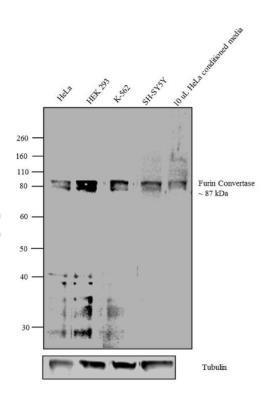
- Western Blot: Furin Antibody [NB100-1903] - Analysis was performed on membrane enriched extracts (30 ug lysate) of HeLa (Lane 1), HEK 293 (Lane 2), K-562 (Lane 3), SH-SY5Y (Lane 4) and 10uL conditioned media from HeLa cell line (Lane 5).
Supportive validation
- Submitted by
- Novus Biologicals (provider)
- Main image
- Experimental details
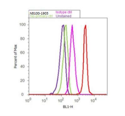
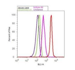
- Flow Cytometry: Furin Antibody [NB100-1903] - Flow cytometry analysis of Furin Convertase was done on HeLa cells. Cells were fixed with 70% ethanol for 10 minutes, permeabilized with 0.25% Triton (R) X-100 for 20 minutes, and blocked with 5% BSA for 30 minutes at room temperature. Cells were labeled with Furin Convertase Rabbit Polyclonal Antibody or with rabbit isotype control (pink histogram) at 3-5 ug/million cells in 2.5% BSA. After incubation at room temperature for 2 hours, the cells were labeled with Alexa Fluor (R) 488 Goat Anti-Rabbit Secondary Antibody (A11008) at a dilution of 1:400 for 30 minutes at room temperature. The representative 10, 000 cells were acquired and analyzed for each sample using an Attune (R) Acoustic Focusing Cytometer. The purple histogram represents unstained control cells and the green histogram represents no-primary-antibody control.