 Explore
 Validate
 Learn
Learn Western blot
 Immunohistochemistry
Antibody data
- Antibody Data
- Antigen structure
- References [0]
- Comments [0]
- Validations
- Immunohistochemistry [1]
- Flow cytometry [2]
Submit
Validation data
Reference
Comment
Report error
- Product number
- GTX22862 - Provider product page

- Provider
- GeneTex
- Proper citation
- GeneTex Cat#GTX22862, RRID:AB_384882
- Product name
- Cav1.1 antibody [1A]
- Antibody type
- Monoclonal
- Reactivity
- Human, Mouse, Rat, Guinea Pig, Rabbit
- Host
- Mouse
No comments: Submit comment
Supportive validation
- Submitted by
- GeneTex (provider)
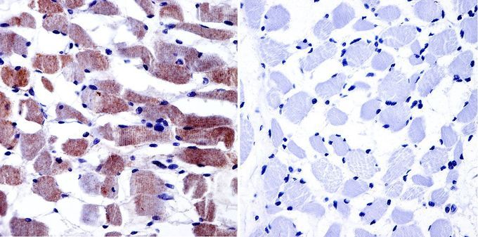
- Main image
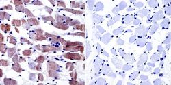
- Experimental details
- Immunohistochemistry was performed on normal biopsies of deparaffinized human skeletal muscle tissue. To expose target proteins, heat induced antigen retrieval was performed using 10mM sodium citrate (pH6.0) buffer, microwaved for 8-15 minutes. Following antigen retrieval tissues were blocked in 3% BSA-PBS for 30 minutes at room temperature. Tissues were then probed at a dilution of 1:20 with or without Cav1.1 antibody [1A] overnight at 4¢XC in a humidified chamber. Tissues were washed extensively with PBST and endogenous peroxidase activity was quenched with a peroxidase suppressor. Detection was performed using a biotin-conjugated secondary antibody and SA-HRP, followed by colorimetric detection using DAB. Tissues were counterstained with hematoxylin and prepped for mounting.
Supportive validation
- Submitted by
- GeneTex (provider)
- Main image
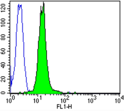
- Experimental details
- Flow cytometry analysis of Dihydropyridine Receptor alpha-2 showing positive staining in the membrane and cytoplasm of C6 cells compared to an isotype control (blue). Cells were harvested, adjusted to a concentration of 1-5x10^6 cells/ml, fixed with 2% paraformaldehyde and washed with PBS. Cells were blocked with a 2% solution of BSA-PBS for 30 min at room temperature and incubated with a Dihydropyridine Receptor alpha-2 monoclonal antibody (GTX22864) at a dilution of 1:100 for 60 min at room temperature. Cells were then incubated for 40 min at room temperature in the dark using a Dylight 488-conjugated goat anti-mouse IgG (H+L) secondary antibody and re-suspended in PBS for FACS analysis.
- Validation comment
- FACS

- Submitted by
- GeneTex (provider)
- Main image
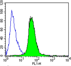
- Experimental details
- Flow cytometry analysis of Dihydropyridine Receptor alpha-2 showing positive staining in the membrane and cytoplasm of SH-SY5Y cells compared to an isotype control (blue). Cells were harvested, adjusted to a concentration of 1-5x10^6 cells/ml, fixed with 2% paraformaldehyde and washed with PBS. Cells were blocked with a 2% solution of BSA-PBS for 30 min at room temperature and incubated with a Dihydropyridine Receptor alpha-2 monoclonal antibody (GTX22864) at a dilution of 1:100 for 60 min at room temperature. Cells were then incubated for 40 min at room temperature in the dark using a Dylight 488-conjugated goat anti-mouse IgG (H+L) secondary antibody and re-suspended in PBS for FACS analysis.
- Validation comment
- FACS
