Explore
 Validate
 Learn
Learn Western blot
Antibody data
- Antibody Data
- Antigen structure
- References [1]
- Comments [0]
- Validations
- Western blot [4]
- Immunohistochemistry [1]
- Other assay [1]
Submit
Validation data
Reference
Comment
Report error
- Product number
- PA5-44189 - Provider product page

- Provider
- Invitrogen Antibodies
- Product name
- GCLC Polyclonal Antibody
- Antibody type
- Polyclonal
- Antigen
- Synthetic peptide
- Description
- Peptide sequence: VLETLQEKGE RTNPNHPTLW RPEYGSYMIE GTPGQPYGGT MSEFNTVEAN
- Concentration
- 0.5 mg/mL
Submitted references Defective protein repair under methionine sulfoxide A deletion drives autophagy and ARE-dependent gene transcription.
Pennington SM, Klutho PR, Xie L, Broadhurst K, Koval OM, McCormick ML, Spitz DR, Grumbach IM
Redox biology 2018 Jun;16:401-413
Redox biology 2018 Jun;16:401-413
No comments: Submit comment
Supportive validation
- Submitted by
- Invitrogen Antibodies (provider)
- Main image
- Experimental details
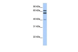
- Western blot analysis of mouse heart cell lysate using an anti-GCLC polyclonal antibody (Product # PA5-44189). Primary Antibody Dilution: 1:1000; Secondary Antibody: Anti-rabbit HRP; Secondary Antibody Dilution: 1:10000. LANE 1) 40µg mouse heart lysate; LANE 2) 40µg mouse heart lysate; LANE 3) 40µg mouse heart lysate; LANE 4) 40µg mouse heart lysate; LANE 5) 40µg mouse heart lysate.
- Submitted by
- Invitrogen Antibodies (provider)
- Main image
- Experimental details
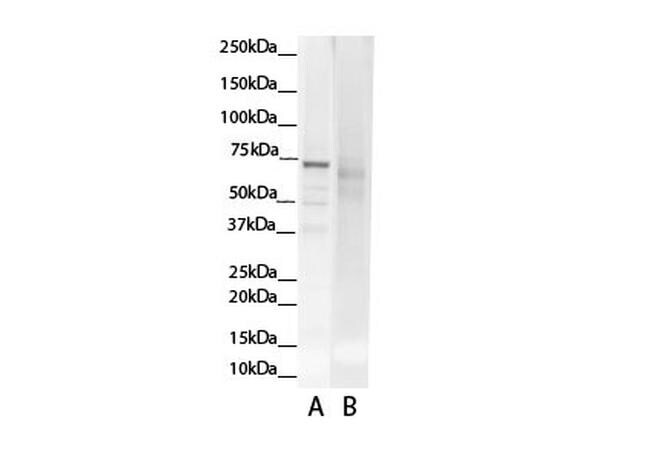
- Western blot analysis of human liver cells using an anti-GCLC polyclonal antibody (Product # PA5-44189).

- Submitted by
- Invitrogen Antibodies (provider)
- Main image

- Experimental details
- Western blot analysis of human liver cells using an anti-GCLC polyclonal antibody (Product # PA5-44189).
- Submitted by
- Invitrogen Antibodies (provider)
- Main image
- Experimental details
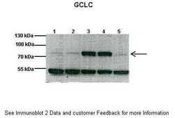
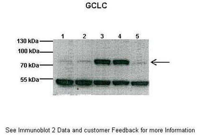
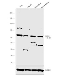
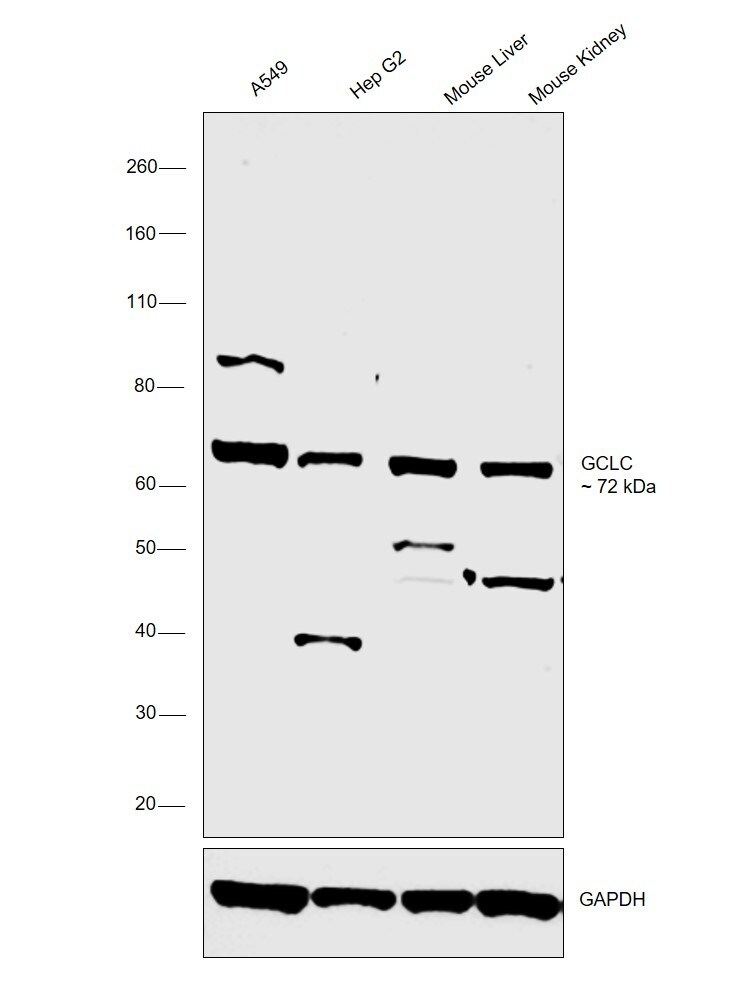
- Western blot was performed using Anti-GCLC Polyclonal Antibody (Product # PA5-44189) and a 72 kDa band corresponding to GCLC was observed across all the cell lines and tissue tested. Whole cell extracts (30 µg lysate) of A549 (Lane 1), Hep G2 (Lane 2), tissue extracts of Mouse Liver (Lane 3) and Mouse Kidney (Lane 4) were electrophoresed using Novex® NuPAGE® 4-12% Bis-Tris Protein Gel (Product # NP0321BOX). Resolved proteins were then transferred onto a nitrocellulose membrane (Product # IB23001) by iBlot® 2 Dry Blotting System (Product # IB21001). The blot was probed with the primary antibody (0.4µg/ml) and detected by chemiluminescence with Goat anti-Rabbit IgG (H+L), Superclonal™ Recombinant Secondary Antibody, HRP (Product # A27036, 1:4000 dilution) using the iBright FL 1000 (Product # A32752). Chemiluminescent detection was performed using Novex® ECL Chemiluminescent Substrate Reagent Kit (Product # WP20005).
Supportive validation
- Submitted by
- Invitrogen Antibodies (provider)
- Main image
- Experimental details
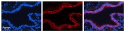
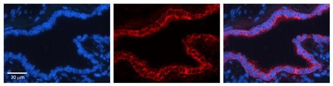
- Immunohistochemistry (paraffin-embedded) analysis of human bronchial epithelial tissue using an anti-GCLC polyclonal antibody (Product # PA5-44189).; Secondary Antibody: Donkey anti-Rabbit-Cy3.
Supportive validation
- Submitted by
- Invitrogen Antibodies (provider)
- Main image

- Experimental details
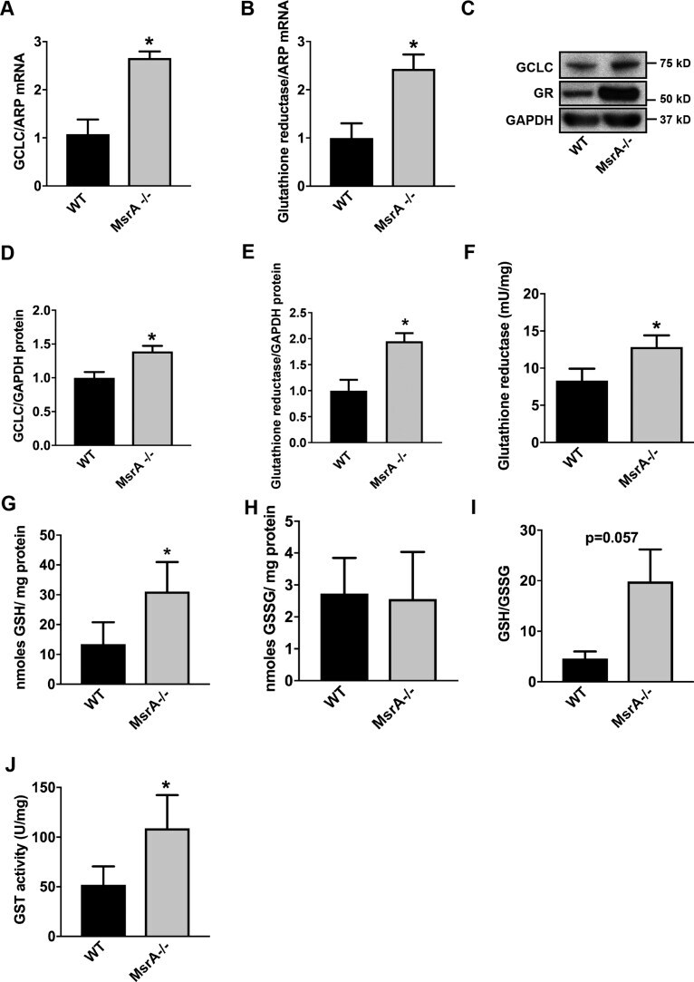
- Fig. 5 Expression and activity of Nrf2-regulated genes are elevated under MsrA deletion. (A, B) GCLC (A) and GR (B) mRNA levels in MsrA-/- and WT VSMC by qRT-PCR, normalized to ARP; n = 3 biological replicates. (C-E) Representative immunoblot (C) and summary data for GCLC (D) and GR (E) protein levels in whole cell lysates from MsrA-/- and WT VSMC. Data were normalized to GAPDH; n = 6-7 biological replicates. (F) GR activity, normalized to total protein concentration; n = 4 biological replicates. (G, H) Levels of reduced glutathione (GSH, G) and oxidized glutathione (GSSG, H), normalized to total protein; n = 4 biological replicates. (I) Ratio of reduced/oxidized glutathione. (J) Glutathione transferase (GST) activity, normalized to total protein; n = 7 biological replicates. * p < 0.05 versus WT by two-tailed t-test. Fig. 5