 Explore
 Validate
 Learn
Learn Western blot
 Immunoprecipitation
Antibody data
- Antibody Data
- Antigen structure
- References [0]
- Comments [0]
- Validations
- Western blot [1]
- Immunocytochemistry [3]
- Immunohistochemistry [1]
Submit
Validation data
Reference
Comment
Report error
- Product number
- MA1-23185 - Provider product page

- Provider
- Invitrogen Antibodies
- Product name
- CENPF Monoclonal Antibody (14C10 1D8)
- Antibody type
- Monoclonal
- Antigen
- Other
- Description
- Recommended positive controls: HeLa (G2 phase). Store product as a concentrated solution. Centrifuge briefly prior to opening the vial.
- Reactivity
- Human
- Host
- Mouse
- Isotype
- IgG
- Antibody clone number
- 14C10 1D8
- Vial size
- 100 µL
- Concentration
- 1 mg/mL
- Storage
- Store at 4°C short term. For long term storage, store at -20°C, avoiding freeze/thaw cycles.
No comments: Submit comment
Supportive validation
- Submitted by
- Invitrogen Antibodies (provider)
- Main image

- Experimental details
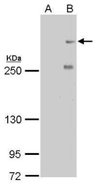
- CENPF Monoclonal Antibody (14C10 1D8) detects CENPF protein by western blot analysis. A. 30 µg HeLa whole cell lysate/extract (untreated). B. 30 µg HeLa whole cell lysate/extract (100 ng/mL Nocodazole treatment for 24hr).5% SDS-PAGE. CENPF Monoclonal Antibody (14C10 1D8) (Product # MA1-23185) dilution: 1:500. The HRP-conjugated anti-mouse IgG antibody was used to detect the primary antibody.
Supportive validation
- Submitted by
- Invitrogen Antibodies (provider)
- Main image

- Experimental details
- Immunofluorescent analysis of Mitosin in U2OS cells fixed by 4% PFA. Cells were costained using a Mitosin monoclonal antibody (Product # MA1-23185) at a dilution of 1:500 and an Alpha-Tubulin polyclonal antibody (Product # PA5-29444) (Green) at a dilution of 1:500. Chromosomes are stained with DAPI (blue). Scale bar, 5 um.

- Submitted by
- Invitrogen Antibodies (provider)
- Main image

- Experimental details
- Confocal immunofluorescence staining (Olympus FV10i) of Mitosin. U2OS cells were fixed by 4% PFA and costained with Mitosin 14C10 1D8 AB (Red; CENPF Monoclonal Antibody (14C10 1D8) (Product # MA1-23185); 1:500 Ab dilution) and alpha-tubulin rabbit polyclonal AB (Green; cat# TUBA1A Polyclonal Antibody (Product # PA5-29444); 1:500 Ab dilution ), a spindle marker. DAPI (blue), chromosomes. Scale bar, 5 um.

- Submitted by
- Invitrogen Antibodies (provider)
- Main image

- Experimental details
- Immunofluorescence analysis of CENPF was performed using HeLa cells. The cells were fixed with 4% paraformaldehyde for 10 minutes, permeabilized with 0.1% Triton™ X-100 for 15 minutes, and blocked with 2% BSA for 1 hour at room temperature. The cells were labeled with CENPF Monoclonal Antibody (Product # MA1-23185) at 1:200 dilution in 0.1% BSA and incubated overnight at 4 degree and then labeled with Goat anti-Mouse IgG (H+L) Cross-Adsorbed Secondary Antibody, Alexa Fluor 488 (Product # A32723) at a dilution of 1:2000 for 45 minutes at room temperature (Panel a: green) in HeLa cells. Nuclei (Panel b: blue) were stained with ProLong™ Diamond Antifade Mountant with DAPI (Product # P36962). F-actin (Panel c: red) was stained with Rhodamine Phalloidin (Product # R415, 1:300). Panel d represents the merged image of HeLa cells showing strong nuclear and weak cytoplasmic localization for CENPF. Panel e represents control cells with no primary antibody to assess background. The images were captured at 60X magnification.

Supportive validation
- Submitted by
- Invitrogen Antibodies (provider)
- Main image

- Experimental details
- CENPF Monoclonal Antibody (14C10 1D8) detects CENPF protein at nucleus on BT483 xenograft by immunohistochemical analysis. Sample: Paraffin-embedded BT483 xenograft. CENPF Monoclonal Antibody (14C10 1D8) (Product # MA1-23185) dilution: 1:200. Antigen Retrieval: EDTA based buffer, pH 8.0, 15 min.
