Explore
 Validate
 Learn
Learn Western blot
Antibody data
- Antibody Data
- Antigen structure
- References [0]
- Comments [0]
- Validations
- Western blot [1]
- Flow cytometry [2]
Submit
Validation data
Reference
Comment
Report error
- Product number
- 10-7590-25 - Provider product page

- Provider
- ABEOMICS Inc.
- Product name
- Anti-Spectrin beta 3 Antibody
- Antibody type
- Monoclonal
- Description
- Beta-III spectrin is a protein encoded by the gene SPTBN2, spans 2390 amino acids and consists of an amino-terminal actin binding domain (ABD), a central region containing seventeen spectrin repeat domains, and a carboxy-terminal pleckstrin homology domain. It is critical for the correct development and maintenance of Purkinje cell dendritic structure. Beta-III spectrin is expressed predominantly in the brain and is enriched in cerebellar Purkinje cells. In addition, Beta-III spectrin participates in endomembrane trafficking through its interaction with the actin related protein, ARP1. The functional unit of Beta-III spectrin is considered to be a tetrameric complex composed of two Beta-spectrin subunits and two Alpha-II-spectrin subunits. Mutations in the gene encoding Beta-III spectrin give rise to spinocerebellar ataxia type 5, a neurodegenerative disease characterized by progressive thinning of the molecular layer, loss of Purkinje cells and increasing motor deficits.
- Reactivity
- Human
- Host
- Mouse
- Conjugate
- Unconjugated
- Antigen sequence
A partial length recombinant protei
n  (a.a 1280-1450) of Spectrin beta
 3 was used as the immunogen for th
is antibody.- Isotype
- IgG
- Antibody clone number
- ABM5A16
- Vial size
- 100 µg
- Concentration
- 0.5 mg/ml
- Storage
- Store the antibody at 4°C, stable for 6 months. For long-term storage, store at -20°C. Avoid repeat freez thawing
No comments: Submit comment
Supportive validation
- Submitted by
- ABEOMICS Inc. (provider)
- Main image

- Experimental details
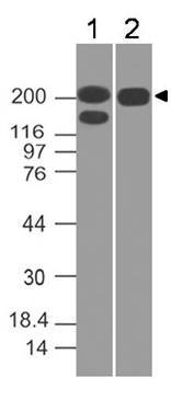
- Western blot analysis of Spectrin beta 3. Anti-Spectrin beta 3 antibody (Clone: ABM5A16) was used at 0.1 µg/ml on h Kidney and h Brain lysates.
- Protocol
- Protocol
Supportive validation
- Submitted by
- ABEOMICS Inc. (provider)
- Main image

- Experimental details
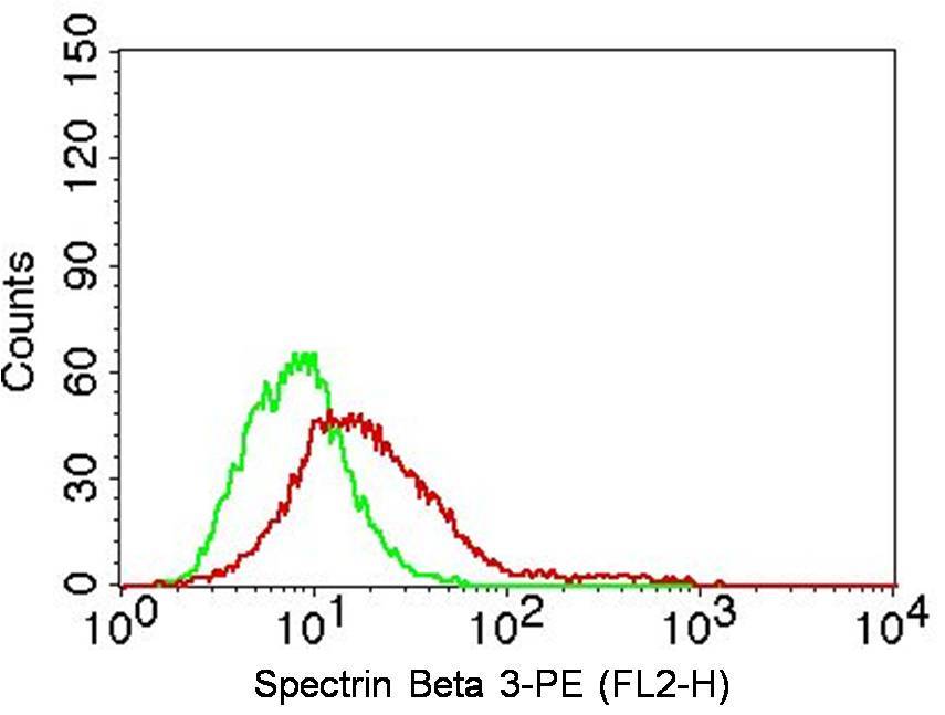
- Intracellular flow analysis of human Spectrin beta 3 in A431 cells using 2 µg/10^6 cells of antibody (Clone: ABM5A16). Green represents isotype control; red represents anti-Spectrin beta 3 antibody. Goat anti-mouse PE conjugate was used as secondary antibody. (Cells were fixed with 4% paraformaldehyde for 10 min and washed with PBS by centrifuging at 1100 for 5 min followed by permeabilization for 20 min and washed again as mentioned above. Then cell were incubated with primary antibody for 45 min. and after washing the cells twice in PBS, incubated with conjugated secondary antibody for 30 min. Data acquisition was done after washing twice with PBS as mentioned above).
- Protocol
- Protocol
- Submitted by
- ABEOMICS Inc. (provider)
- Main image

- Experimental details
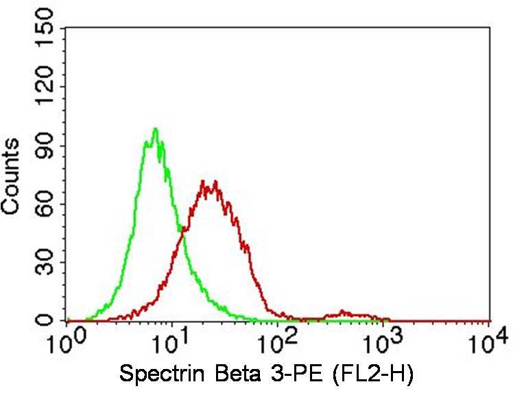
- Intracellular flow analysis of human Spectrin beta 3 in HeLa cells using 2 µg/10^6 cells of antibody (Clone: ABM5A16). Green represents isotype control; red represents anti-Spectrin beta 3 antibody. Goat anti-mouse PE conjugate was used as secondary antibody. (Cells were fixed with 4% paraformaldehyde for 10 min and washed with PBS by centrifuging at 1100 for 5 min followed by permeabilization for 20 min and washed again as mentioned above. Then cell were incubated with primary antibody for 45 min. and after washing the cells twice in PBS, incubated with conjugated secondary antibody for 30 min. Data acquisition was done after washing twice with PBS as mentioned above).
- Protocol
- Protocol