 Explore
 Validate
 Learn
Learn Western blot
Antibody data
- Antibody Data
- Antigen structure
- References [4]
- Comments [0]
- Validations
- Western blot [2]
- Immunohistochemistry [2]
Submit
Validation data
Reference
Comment
Report error
- Product number
- AF-258-PB - Provider product page

- Provider
- R&D Systems
- Product name
- Human Midkine Antibody
- Antibody type
- Polyclonal
- Description
- Antigen Affinity-purified. Detects human Midkine in direct ELISAs and Western blots. In direct ELISAs, approximately 10% cross-reactivity with recombinant mouse Midkine is observed, and less than 1% cross-reactivity with recombinant human Pleiotrophin is observed.
- Reactivity
- Human
- Host
- Goat
- Conjugate
- Unconjugated
- Antigen sequence
P21741- Isotype
- IgG
- Vial size
- 100 ug
- Concentration
- LYOPH
- Storage
- Use a manual defrost freezer and avoid repeated freeze-thaw cycles. 12 months from date of receipt, -20 to -70 °C as supplied. 1 month, 2 to 8 °C under sterile conditions after reconstitution. 6 months, -20 to -70 °C under sterile conditions after reconstitution.
Submitted references Role of heparin binding growth factors in nigrostriatal dopamine system development and Parkinson's disease.
Regulation of MDK expression in human cancer cells modulates sensitivities to various anticancer drugs: MDK overexpression confers to a multi-drug resistance.
Neuritogenic activity of chondroitin/dermatan sulfate hybrid chains of embryonic pig brain and their mimicry from shark liver. Involvement of the pleiotrophin and hepatocyte growth factor signaling pathways.
The anti-HIV cytokine midkine binds the cell surface-expressed nucleolin as a low affinity receptor.
Marchionini DM, Lehrmann E, Chu Y, He B, Sortwell CE, Becker KG, Freed WJ, Kordower JH, Collier TJ
Brain research 2007 May 25;1147:77-88
Brain research 2007 May 25;1147:77-88
Regulation of MDK expression in human cancer cells modulates sensitivities to various anticancer drugs: MDK overexpression confers to a multi-drug resistance.
Kang HC, Kim IJ, Park HW, Jang SG, Ahn SA, Yoon SN, Chang HJ, Yoo BC, Park JG
Cancer letters 2007 Mar 8;247(1):40-7
Cancer letters 2007 Mar 8;247(1):40-7
Neuritogenic activity of chondroitin/dermatan sulfate hybrid chains of embryonic pig brain and their mimicry from shark liver. Involvement of the pleiotrophin and hepatocyte growth factor signaling pathways.
Li F, Shetty AK, Sugahara K
The Journal of biological chemistry 2007 Feb 2;282(5):2956-66
The Journal of biological chemistry 2007 Feb 2;282(5):2956-66
The anti-HIV cytokine midkine binds the cell surface-expressed nucleolin as a low affinity receptor.
Said EA, Krust B, Nisole S, Svab J, Briand JP, Hovanessian AG
The Journal of biological chemistry 2002 Oct 4;277(40):37492-502
The Journal of biological chemistry 2002 Oct 4;277(40):37492-502
No comments: Submit comment
Supportive validation
- Submitted by
- R&D Systems (provider)
- Main image

- Experimental details
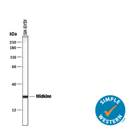
- Detection of Human Midkine by Western Blot. Western blot shows lysates of SH-SY5Y human neuroblastoma cell line. PVDF membrane was probed with 2 µg/mL of Goat Anti-Human Midkine Antigen Affinity-purified Polyclonal Antibody (Catalog # AF-258-PB) followed by HRP-conjugated Anti-Goat IgG Secondary Antibody (Catalog # HAF017). A specific band was detected for Midkine at approximately 17 kDa (as indicated). This experiment was conducted under reducing conditions and using Immunoblot Buffer Group 1.

- Submitted by
- R&D Systems (provider)
- Main image
- Experimental details
- Detection of Human Midkine by Simple WesternTM. Simple Western lane view shows lysates of SH-SY5Y human neuroblastoma cell line, loaded at 0.2 mg/mL. A specific band was detected for Midkine at approximately 26 kDa (as indicated) using 100 µg/mL of Goat Anti-Human Midkine Antigen Affinity-purified Polyclonal Antibody (Catalog # AF-258-PB) followed by 1:50 dilution of HRP-conjugated Anti-Goat IgG Secondary Antibody (Catalog # HAF109). This experiment was conducted under reducing conditions and using the 12-230 kDa separation system.

Supportive validation
- Submitted by
- R&D Systems (provider)
- Main image

- Experimental details
- Midkine in Human Prostate. Midkine was detected in immersion fixed paraffin-embedded sections of human prostate using Goat Anti-Human Midkine Antigen Affinity-purified Polyclonal Antibody (Catalog # AF-258-PB) at 10 µg/mL overnight at 4 °C. Before incubation with the primary antibody, tissue was subjected to heat-induced epitope retrieval using Antigen Retrieval Reagent-Basic (Catalog # CTS013). Tissue was stained using the Anti-Goat HRP-DAB Cell & Tissue Staining Kit (brown; Catalog # CTS008) and counterstained with hematoxylin (blue). Specific staining was localized to stromal cell cytoplasm. View our protocol for Chromogenic IHC Staining of Paraffin-embedded Tissue Sections.

- Submitted by
- R&D Systems (provider)
- Main image
- Experimental details
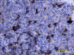
- Midkine in Human Ovarian Array. Midkine was detected in immersion fixed paraffin-embedded sections of human ovarian array using Goat Anti-Human Midkine Antigen Affinity-purified Polyclonal Antibody (Catalog # AF-258-PB) at 10 µg/mL overnight at 4 °C. Tissue was stained using the Anti-Goat HRP-DAB Cell & Tissue Staining Kit (brown; Catalog # CTS008) and counterstained with hematoxylin (blue). View our protocol for Chromogenic IHC Staining of Paraffin-embedded Tissue Sections.
