 Explore
 Validate
 Learn
Learn Western blot
Antibody data
- Antibody Data
- Antigen structure
- References [4]
- Comments [0]
- Validations
- Western blot [1]
- Immunohistochemistry [1]
Submit
Validation data
Reference
Comment
Report error
- Product number
- sc-540 - Provider product page

- Provider
- Santa Cruz Biotechnology
- Proper citation
- Santa Cruz Biotechnology Cat#sc-540, RRID:AB_632567
- Product name
- Anti-TTK
- Antibody type
- Polyclonal (Antigen purified)
- Antigen
- Synthetic peptide
- Reactivity
- Human
- Host
- Rabbit
Submitted references TTK/hMps1 mediates the p53-dependent postmitotic checkpoint by phosphorylating p53 at Thr18.
Chk2-dependent phosphorylation of XRCC1 in the DNA damage response promotes base excision repair.
Ablation of the spindle assembly checkpoint by a compound targeting Mps1.
Dynamic distribution of TTK in HeLa cells: insights from an ultrastructural study
Huang YF, Chang MD, Shieh SY
Molecular and cellular biology 2009 Jun;29(11):2935-44
Molecular and cellular biology 2009 Jun;29(11):2935-44
Chk2-dependent phosphorylation of XRCC1 in the DNA damage response promotes base excision repair.
Chou WC, Wang HC, Wong FH, Ding SL, Wu PE, Shieh SY, Shen CY
The EMBO journal 2008 Dec 3;27(23):3140-50
The EMBO journal 2008 Dec 3;27(23):3140-50
Ablation of the spindle assembly checkpoint by a compound targeting Mps1.
Schmidt M, Budirahardja Y, Klompmaker R, Medema RH
EMBO reports 2005 Sep;6(9):866-72
EMBO reports 2005 Sep;6(9):866-72
Dynamic distribution of TTK in HeLa cells: insights from an ultrastructural study
Zhen DOU, Akira SAWAGECHI, Jie ZHANG, Hong LUO, Lawrence BRAKO, Xue Biao YAO
Cell Research 2003 Dec;13(6):443-449
Cell Research 2003 Dec;13(6):443-449
No comments: Submit comment
Supportive validation
- Submitted by
- per
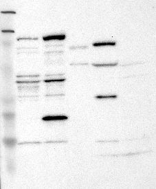
- Main image

- Experimental details
- Western blot analysis of antibody specificity using a routine panel composed of IgG/HSA-depleted human plasma and protein lysates from selected human tissues and cell lines.
- Validation comment
- Band of predicted size in kDa (+/-20%) with additional bands present.
- Primary Ab dilution
- 1:500
- Secondary Ab dilution
- 1:3000
- Lane 1
- Marker [kDa]: 219, 111, 83, 48, 32, 26, 17
- Lane 2
- RT-4
- Lane 3
- U-251MG sp
- Lane 4
- Human Plasma
- Lane 5
- Liver
- Lane 6
- Tonsil
- Theoretical target weight
- [kDa] 97
Supportive validation
- Submitted by
- per
- Main image
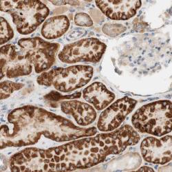
- Experimental details
- Immunohistochemical staining of human kidney shows strong cytoplasmic positivity in cells of tubules.
- Validation comment
- Staining pattern consistent with experimental and/or bioinformatic data.
