 Explore
 Validate
 Learn
Learn Western blot
Antibody data
- Antibody Data
- Antigen structure
- References [1]
- Comments [0]
- Validations
- Western blot [1]
- Immunohistochemistry [8]
- Flow cytometry [2]
Submit
Validation data
Reference
Comment
Report error
- Product number
- TA504017 - Provider product page

- Provider
- OriGene
- Proper citation
- OriGene Cat#TA504017, RRID:AB_11125011
- Product name
- TIMP2 mouse monoclonal antibody, clone OTI1A6 (formerly 1A6)
- Antibody type
- Monoclonal
- Description
- TIMP2 mouse monoclonal antibody, clone OTI1A6 (formerly 1A6)
- Reactivity
- Canine
- Host
- Mouse
- Conjugate
- Unconjugated
- Epitope
- TIMP2
- Isotype
- IgG
- Antibody clone number
- OTI1A6
- Vial size
- 100 µl
- Concentration
- NULL
Submitted references Statins' Withdrawal Induces Atherosclerotic Plaque Destabilization in Animal Model-A "Rebound" Stimulation of Inflammation.
Stasinopoulou M, Kadoglou NPE, Christodoulou E, Paronis E, Kostomitsopoulos NG, Valsami G, Liapis CD, Kakisis J
Journal of cardiovascular pharmacology and therapeutics 2019 Jul;24(4):377-386
Journal of cardiovascular pharmacology and therapeutics 2019 Jul;24(4):377-386
No comments: Submit comment
Supportive validation
- Submitted by
- OriGene (provider)
- Main image

- Experimental details
- HEK293T cells were transfected with the pCMV6-ENTRY control (Left lane) or pCMV6-ENTRY TIMP2 (RC209796, Right lane) cDNA for 48 hrs and lysed. Equivalent amounts of cell lysates (5 ug per lane) were separated by SDS-PAGE and immunoblotted with anti-TIMP2.
- Validation comment
- WB

Supportive validation
- Submitted by
- OriGene (provider)
- Main image
- Experimental details
- Immunohistochemical staining of paraffin-embedded Adenocarcinoma of Human breast tissue using anti-TIMP2 mouse monoclonal antibody. (Heat-induced epitope retrieval by 10mM citric buffer, pH6.0, 100C for 10min, TA504017)
- Validation comment
- IHC

- Submitted by
- OriGene (provider)
- Main image

- Experimental details
- Immunohistochemical staining of paraffin-embedded Human endometrium tissue within the normal limits using anti-TIMP2 mouse monoclonal antibody. (Heat-induced epitope retrieval by 10mM citric buffer, pH6.0, 100C for 10min, TA504017)
- Validation comment
- IHC

- Submitted by
- OriGene (provider)
- Main image

- Experimental details
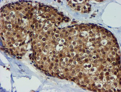
- Immunohistochemical staining of paraffin-embedded Human breast tissue within the normal limits using anti-TIMP2 mouse monoclonal antibody. (Heat-induced epitope retrieval by 10mM citric buffer, pH6.0, 100C for 10min, TA504017)
- Validation comment
- IHC

- Submitted by
- OriGene (provider)
- Main image

- Experimental details
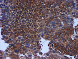
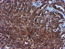
- Immunohistochemical staining of paraffin-embedded Carcinoma of Human bladder tissue using anti-TIMP2 mouse monoclonal antibody. (Heat-induced epitope retrieval by 10mM citric buffer, pH6.0, 100C for 10min, TA504017)
- Validation comment
- IHC
- Submitted by
- OriGene (provider)
- Main image
- Experimental details
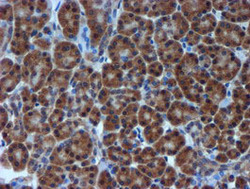
- Immunohistochemical staining of paraffin-embedded Human pancreas tissue within the normal limits using anti-TIMP2 mouse monoclonal antibody. (Heat-induced epitope retrieval by 10mM citric buffer, pH6.0, 100C for 10min, TA504017)
- Validation comment
- IHC

- Submitted by
- OriGene (provider)
- Main image

- Experimental details
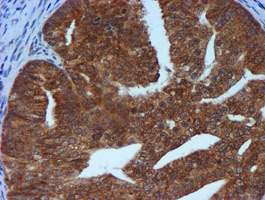
- Immunohistochemical staining of paraffin-embedded Carcinoma of Human lung tissue using anti-TIMP2 mouse monoclonal antibody. (Heat-induced epitope retrieval by 10mM citric buffer, pH6.0, 100C for 10min, TA504017)
- Validation comment
- IHC

- Submitted by
- OriGene (provider)
- Main image

- Experimental details
- Immunohistochemical staining of paraffin-embedded Adenocarcinoma of Human endometrium tissue using anti-TIMP2 mouse monoclonal antibody. (Heat-induced epitope retrieval by 10mM citric buffer, pH6.0, 100C for 10min, TA504017)
- Validation comment
- IHC
- Submitted by
- OriGene (provider)
- Main image

- Experimental details
- Immunohistochemical staining of paraffin-embedded Adenocarcinoma of Human ovary tissue using anti-TIMP2 mouse monoclonal antibody. (Heat-induced epitope retrieval by 10mM citric buffer, pH6.0, 100C for 10min, TA504017)
- Validation comment
- IHC
Supportive validation
- Submitted by
- OriGene (provider)
- Main image

- Experimental details
- Flow cytometric Analysis of Jurkat cells, using anti-TIMP2 antibody(TA504017),(Red), compared to a nonspecific negative control antibody,(Blue).
- Validation comment
- FC

- Submitted by
- OriGene (provider)
- Main image

- Experimental details
- HEK293T cells transfected with either RC209796 overexpress plasmid(Red) or empty vector control plasmid(Blue) were immunostained by anti-TIMP2 antibody(TA504017), and then analyzed by flow cytometry.
- Validation comment
- FC
