 Explore
 Validate
 Learn
Learn Western blot
Antibody data
- Antibody Data
- Antigen structure
- References [1]
- Comments [0]
- Validations
- Western blot [3]
- Immunocytochemistry [3]
- Immunohistochemistry [6]
Submit
Validation data
Reference
Comment
Report error
- Product number
- MA5-26269 - Provider product page

- Provider
- Invitrogen Antibodies
- Product name
- PMS2 Monoclonal Antibody (OTI2G5)
- Antibody type
- Monoclonal
- Antigen
- Recombinant full-length protein
- Reactivity
- Human
- Host
- Mouse
- Isotype
- IgG
- Antibody clone number
- OTI2G5
- Vial size
- 100 µL
- Concentration
- 1 mg/mL
- Storage
- -20° C, Avoid Freeze/Thaw Cycles
Submitted references Focal amplifications are associated with chromothripsis events and diverse prognoses in gastric cardia adenocarcinoma.
Zhao XK, Xing P, Song X, Zhao M, Zhao L, Dang Y, Lei LL, Xu RH, Han WL, Wang PP, Yang MM, Hu JF, Zhong K, Zhou FY, Han XN, Meng CL, Ji JJ, Chen X, Wang LD
Nature communications 2021 Nov 11;12(1):6489
Nature communications 2021 Nov 11;12(1):6489
No comments: Submit comment
Supportive validation
- Submitted by
- Invitrogen Antibodies (provider)
- Main image

- Experimental details
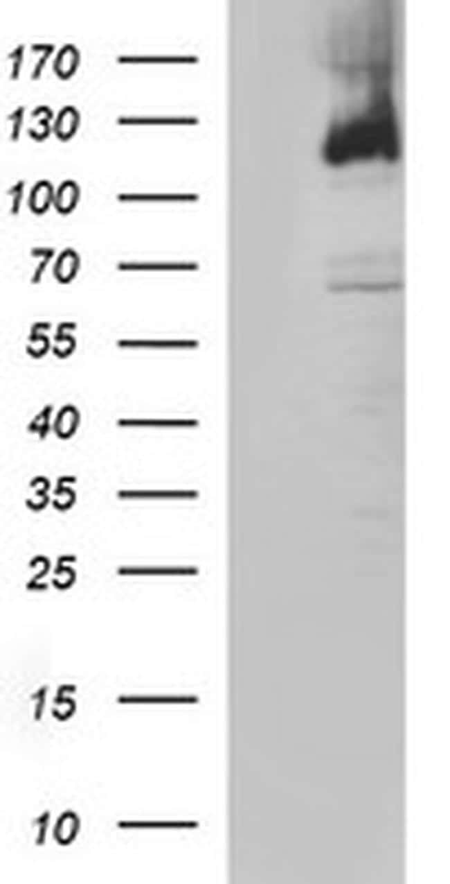
- Western blot analysis of PMS2 in HEK293T cells in untransfected (Left lane) and transfected (Right lane) samples using 5 µg per lane. The samples were separated by SDS-PAGE and probed with PMS2 (Product # MA5-26269) monoclonal antibody.
- Submitted by
- Invitrogen Antibodies (provider)
- Main image

- Experimental details
- Western blot analysis of PMS2 in HEK293T cells in untransfected (Left lane) and transfected (Right lane) samples using 5 µg per lane. The samples were separated by SDS-PAGE and probed with PMS2 (Product # MA5-26269) monoclonal antibody.

- Submitted by
- Invitrogen Antibodies (provider)
- Main image

- Experimental details
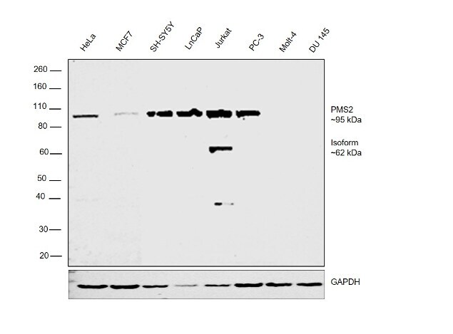
- Western blot was performed using Anti-PMS2 Monoclonal Antibody (OTI2G5) (Product # MA5-26269) and a 95kDa band corresponding to PMS2 was observed in HeLa, MCF7, SH-SY5Y, LNCaP, Jurkat and PC-3 and not in Molt-4 and DU 145. An isoform corresponding to 62kDa was observed only in Jurkat cell line. Whole cell extracts (30 µg lysate) of HeLa (Lane 1), MCF7 (Lane 2), SH-SY5Y (Lane 3), LnCaP (Lane 4), Jurkat (Lane 5), PC-3 (Lane 6), Molt-4 (Lane 7) and DU 145 (Lane 8) were electrophoresed using NuPAGE™ 4-12% Bis-Tris Protein Gel (Product # NP0322BOX). Resolved proteins were then transferred onto a nitrocellulose membrane (Product # IB23001) by iBlot® 2 Dry Blotting System (Product # IB21001). The blot was probed with the primary antibody (1:500 dilution) and detected by chemiluminescence with Goat Anti-Mouse IgG Secondary Antibody, HRP conjugate (Product # A28177, 1:4000 dilution) using the iBright FL 1000 (Product # A32752). Chemiluminescent detection was performed using Novex® ECL Chemiluminescent Substrate Reagent Kit (Product # WP20005).
Supportive validation
- Submitted by
- Invitrogen Antibodies (provider)
- Main image
- Experimental details
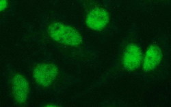
- Immunofluorescent analysis of PMS2 in HeLa cells. Cells were probed with a PMS2 monoclonal antibody (Product # MA5-26269).

- Submitted by
- Invitrogen Antibodies (provider)
- Main image

- Experimental details
- Immunofluorescent analysis of PMS2 in HeLa cells. Cells were probed with a PMS2 monoclonal antibody (Product # MA5-26269).

- Submitted by
- Invitrogen Antibodies (provider)
- Main image

- Experimental details
- Immunofluorescent analysis of PMS2 in HeLa cells. Cells were probed with a PMS2 monoclonal antibody (Product # MA5-26269).

Supportive validation
- Submitted by
- Invitrogen Antibodies (provider)
- Main image

- Experimental details
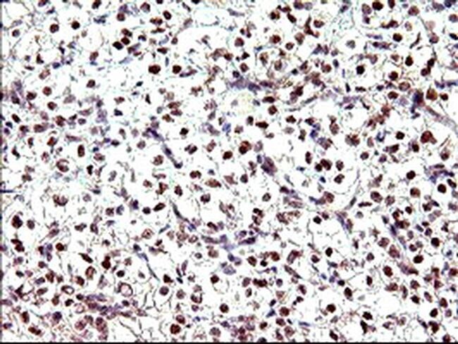
- Immunohistochemistry was performed on paraffin-embedded adenocarcinoma of human breast tissue. To expose target proteins, 10mM citric buffer, pH6.0, 120°C for 3min was used. Following antigen retrieval, tissues were probed with a PMS2 monoclonal antibody (Product # MA5-26269).

- Submitted by
- Invitrogen Antibodies (provider)
- Main image

- Experimental details
- Immunohistochemistry was performed on paraffin-embedded adenocarcinoma of human colon tissue. To expose target proteins, 10mM citric buffer, pH6.0, 120°C for 3min was used. Following antigen retrieval, tissues were probed with a PMS2 monoclonal antibody (Product # MA5-26269).

- Submitted by
- Invitrogen Antibodies (provider)
- Main image

- Experimental details
- Immunohistochemistry was performed on paraffin-embedded human kidney tissue. To expose target proteins, 10mM citric buffer, pH6.0, 120°C for 3min was used. Following antigen retrieval, tissues were probed with a PMS2 monoclonal antibody (Product # MA5-26269).

- Submitted by
- Invitrogen Antibodies (provider)
- Main image

- Experimental details
- Immunohistochemistry was performed on paraffin-embedded carcinoma of human kidney tissue. To expose target proteins, 10mM citric buffer, pH6.0, 120°C for 3min was used. Following antigen retrieval, tissues were probed with a PMS2 monoclonal antibody (Product # MA5-26269).
- Submitted by
- Invitrogen Antibodies (provider)
- Main image

- Experimental details
- Immunohistochemistry was performed on paraffin-embedded adenocarcinoma of human ovary tissue. To expose target proteins, 10mM citric buffer, pH6.0, 120°C for 3min was used. Following antigen retrieval, tissues were probed with a PMS2 monoclonal antibody (Product # MA5-26269).

- Submitted by
- Invitrogen Antibodies (provider)
- Main image

- Experimental details
- Immunohistochemistry was performed on paraffin-embedded carcinoma of human pancreas tissue. To expose target proteins, 10mM citric buffer, pH6.0, 120°C for 3min was used. Following antigen retrieval, tissues were probed with a PMS2 monoclonal antibody (Product # MA5-26269).
